鬼笔环肽-赖氨酸标记 货号5304-AAT Bioquest荧光染料

上海金畔生物科技有限公司代理AAT Bioquest荧光染料全线产品,欢迎访问AAT Bioquest荧光染料官网了解更多信息。

鬼笔环肽-赖氨酸标记

鬼笔环肽-赖氨酸标记

鬼笔环肽-赖氨酸标记    货号5304 货号 5304 存储条件 在零下15度以下保存, 避免光照
规格 100 ug 价格 1164
Ex (nm) Em (nm)
分子量 885.91 溶剂 DMSO
产品详细介绍

简要概述

产品基本信息

货号:5304

产品名称:鬼笔环肽-赖氨酸标记

规格:100ug

储存条件:-15℃避光防潮

保质期:24个月


产品物理化学光谱特性

分子量:885.91

溶剂:DMSO


产品介绍

鬼笔环肽赖氨酸是一种方便的化合物,可用于开发各种鬼笔环肽衍生物,这些衍生物可用于研究细胞结构和其他生物学应用。它易于与胺反应性染料,生物素和其他标签分子(例如NHS酯,异硫氰酸酯和磺酰氯等)反应。鬼笔环肽是一种双环七肽毒素,特异性结合在F-肌动蛋白亚基之间的界面上,将相邻的亚基锁定在一起。鬼笔环肽与肌动蛋白丝的结合比与肌动蛋白单体的结合要紧密得多,从而导致肌动蛋白亚基从丝端解离的速率常数降低,从而通过防止丝解聚而基本稳定了肌动蛋白丝。鬼笔环肽的性质是研究F-肌动蛋白在细胞中分布的有用工具,方法是用荧光类似物标记鬼笔环肽并将其用于染色肌动蛋白丝以进行光学显微镜检查。已经证明,鬼笔环肽的荧光衍生物在定位活细胞或固定细胞中的肌动蛋白丝以及在体外可视化单个肌动蛋白丝方面非常有用。荧光鬼笔环肽衍生物已被用作高分辨率研究肌动蛋白网络的重要工具。 AAT Bioquest为多色成像应用提供了多种具有不同颜色的荧光鬼笔环肽衍生物。金畔生物是AAT Bioquest的中国代理商,为您提供优质的鬼笔环肽-赖氨酸标记。 


参考文献

Hybrid elastomer-plastic microfluidic device as a convenient model for mimicking the blood-brain barrier in vitro.
Authors: Nguyen, Phuoc Quang Huy and Duong, Duong Duy and Kwun, Jun Dae and Lee, Nae Yoon
Journal: Biomedical microdevices (2019): 90

Purinergic Signaling and Aminoglycoside Ototoxicity: The Opposing Roles of P1 (Adenosine) and P2 (ATP) Receptors on Cochlear Hair Cell Survival.
Authors: Lin, Shelly C Y and Thorne, Peter R and Housley, Gary D and Vlajkovic, Srdjan M
Journal: Frontiers in cellular neuroscience (2019): 207

Gross anatomy of the muscle systems and associated innervation of Apatemon cobitidis proterorhini metacercaria (Trematoda: Strigeidea), as visualized by confocal microscopy.
Authors: Stewart, M T and Mousley, A and Koubková, B and Sebelová, S and Marks, N J and Halton, D W
Journal: Parasitology (2003): 273-82

Light and electron microscopic study of changes in the organization of the cortical actin cytoskeleton during chromaffin cell secretion.
Authors: Tchakarov, L E and Zhang, L and Rosé, S D and Tang, R and Trifaró, J M
Journal: The journal of histochemistry and cytochemistry : official journal of the Histochemistry Society (1998): 193-203

Responses induced by tacrine in neuronal and non-neuronal cell lines.
Authors: De Ferrari, G V and von Bernhardi, R and Calderón, F H and Luza, S C and Inestrosa, N C
Journal: Journal of neuroscience research (1998): 435-44

Expression of PAC 1, an epitope associated with two synapse-enriched glycoproteins and a neuronal cytoskeleton-associated polypeptide in developing forebrain neurons.
Authors: Willmott, T and Williamson, T L and Mummery, R and Hawkes, R B and Can, A and Gurd, J W and Gordon-Weeks, P R and Beesley, P W
Journal: Neuroscience (1994): 115-29

Vasoactive amines directly modify endothelial cells to affect polymorphonuclear leukocyte diapedesis in vitro.
Authors: Doukas, J and Shepro, D and Hechtman, H B
Journal: Blood (1987): 1563-9

说明书
鬼笔环肽-赖氨酸标记.pdf

鬼笔环肽-赖氨酸标记 货号5305-AAT Bioquest荧光染料

上海金畔生物科技有限公司代理AAT Bioquest荧光染料全线产品,欢迎访问AAT Bioquest荧光染料官网了解更多信息。

鬼笔环肽-赖氨酸标记

鬼笔环肽-赖氨酸标记

鬼笔环肽-赖氨酸标记    货号5305 货号 5305 存储条件 在零下15度以下保存, 避免光照
规格 1 mg 价格 7956
Ex (nm) Em (nm)
分子量 885.91 溶剂 DMSO
产品详细介绍

简要概述

产品基本信息

货号:5305

产品名称:鬼笔环肽-赖氨酸标记

规格:1mg

储存条件:-15℃避光防潮

保质期:24个月


产品物理化学光谱特性

分子量:885.91

溶剂:DMSO


产品介绍

鬼笔环肽赖氨酸是一种方便的化合物,可用于开发各种鬼笔环肽衍生物,这些衍生物可用于研究细胞结构和其他生物学应用。它易于与胺反应性染料,生物素和其他标签分子(例如NHS酯,异硫氰酸酯和磺酰氯等)反应。鬼笔环肽是一种双环七肽毒素,特异性结合在F-肌动蛋白亚基之间的界面上,将相邻的亚基锁定在一起。鬼笔环肽与肌动蛋白丝的结合比与肌动蛋白单体的结合要紧密得多,从而导致肌动蛋白亚基从丝端解离的速率常数降低,从而通过防止丝解聚而基本稳定了肌动蛋白丝。鬼笔环肽的性质是研究F-肌动蛋白在细胞中分布的有用工具,方法是用荧光类似物标记鬼笔环肽并将其用于染色肌动蛋白丝以进行光学显微镜检查。已经证明,鬼笔环肽的荧光衍生物在定位活细胞或固定细胞中的肌动蛋白丝以及在体外可视化单个肌动蛋白丝方面非常有用。荧光鬼笔环肽衍生物已被用作高分辨率研究肌动蛋白网络的重要工具。 AAT Bioquest为多色成像应用提供了多种具有不同颜色的荧光鬼笔环肽衍生物。金畔生物是AAT Bioquest的中国代理商,为您提供优质的鬼笔环肽-赖氨酸标记。 


参考文献

Hybrid elastomer-plastic microfluidic device as a convenient model for mimicking the blood-brain barrier in vitro.
Authors: Nguyen, Phuoc Quang Huy and Duong, Duong Duy and Kwun, Jun Dae and Lee, Nae Yoon
Journal: Biomedical microdevices (2019): 90

Purinergic Signaling and Aminoglycoside Ototoxicity: The Opposing Roles of P1 (Adenosine) and P2 (ATP) Receptors on Cochlear Hair Cell Survival.
Authors: Lin, Shelly C Y and Thorne, Peter R and Housley, Gary D and Vlajkovic, Srdjan M
Journal: Frontiers in cellular neuroscience (2019): 207

Gross anatomy of the muscle systems and associated innervation of Apatemon cobitidis proterorhini metacercaria (Trematoda: Strigeidea), as visualized by confocal microscopy.
Authors: Stewart, M T and Mousley, A and Koubková, B and Sebelová, S and Marks, N J and Halton, D W
Journal: Parasitology (2003): 273-82

Light and electron microscopic study of changes in the organization of the cortical actin cytoskeleton during chromaffin cell secretion.
Authors: Tchakarov, L E and Zhang, L and Rosé, S D and Tang, R and Trifaró, J M
Journal: The journal of histochemistry and cytochemistry : official journal of the Histochemistry Society (1998): 193-203

Responses induced by tacrine in neuronal and non-neuronal cell lines.
Authors: De Ferrari, G V and von Bernhardi, R and Calderón, F H and Luza, S C and Inestrosa, N C
Journal: Journal of neuroscience research (1998): 435-44

Expression of PAC 1, an epitope associated with two synapse-enriched glycoproteins and a neuronal cytoskeleton-associated polypeptide in developing forebrain neurons.
Authors: Willmott, T and Williamson, T L and Mummery, R and Hawkes, R B and Can, A and Gurd, J W and Gordon-Weeks, P R and Beesley, P W
Journal: Neuroscience (1994): 115-29

Vasoactive amines directly modify endothelial cells to affect polymorphonuclear leukocyte diapedesis in vitro.
Authors: Doukas, J and Shepro, D and Hechtman, H B
Journal: Blood (1987): 1563-9
说明书
鬼笔环肽-赖氨酸标记.pdf

iFluor 665琥珀酰亚胺酯 货号1553-AAT Bioquest荧光染料

上海金畔生物科技有限公司代理AAT Bioquest荧光染料全线产品,欢迎访问AAT Bioquest荧光染料官网了解更多信息。

iFluor 665琥珀酰亚胺酯

iFluor 665琥珀酰亚胺酯

货号 1553 存储条件 在零下15度以下保存, 避免光照
规格 10 mg 价格 15300
Ex (nm) 667 Em (nm) 692
分子量 1210.53 溶剂 DMSO
产品详细介绍

简要概述

产品基本信息

货号:1553

产品名称:iFluor 665琥珀酰亚胺酯

规格:10mg

储存条件:-15℃,避光干燥

保质期:12个月


物理化学光谱特性

分子量:974.20

溶剂:DMSO

Ex(nm):667

Em(nm):692

Abs(nm):661


产品介绍

AAT Bioquest的iFluor 染料针对标记蛋白质(特别是抗体)进行了优化。这些染料是明亮的,耐光的,并且对蛋白质的淬灭作用极小。它们可以被荧光仪器的主要激光线(例如350、405、488、532、555、633和647 nm)激发。 iFluor 665系列的光谱特性类似于AlexaFluor®660(AlexaFluor®是Invitrogen的商标)。 iFluor 665系列的荧光在pH值从3到11时不受pH值的影响。这些光谱特性使该新染料系列成为AlexaFluor®660的出色替代品。在相同条件下,iFluor 665在某些抗体上可提供更强的荧光信号。 iFluor 665 SE相当稳定,并且对蛋白质氨基具有良好的反应性和选择性。

点击查看光谱


参考文献

Recognition of Invasive Prostate Cancer Using a GHRL Polypeptide Probe Targeting GHSR in a Mouse Model In Vivo.
Authors: Ye, Huamao and Yang, Yue and Chen, Rui and Shi, Xiaolei and Fang, Yu and Yang, Jun and Dong, Yuanzhen and Chen, Lili and Xia, Jianghua and Wang, Chao and Yang, Chenghua and Feng, Jun and Wang, Yang and Feng, Xiang and Lü, Chen
Journal: Current pharmaceutical design (2020): 1614-1621

A rapid sensitive, flow cytometry-based method for the detection of Plasmodium vivax-infected blood cells.
Authors: Roobsoong, Wanlapa and Maher, Steven P and Rachaphaew, Nattawan and Barnes, Samantha J and Williamson, Kim C and Sattabongkot, Jetsumon and Adams, John H
Journal: Malaria journal (2014): 55

Reengineering the optical absorption cross-section of photosynthetic reaction centers.
Authors: Dutta, Palash K and Lin, Su and Loskutov, Andrey and Levenberg, Symon and Jun, Daniel and Saer, Rafael and Beatty, J Thomas and Liu, Yan and Yan, Hao and Woodbury, Neal W
Journal: Journal of the American Chemical Society (2014): 4599-604

Comparison of a chimeric anti-carcinoembryonic antigen antibody conjugated with visible or near-infrared fluorescent dyes for imaging pancreatic cancer in orthotopic nude mouse models.
Authors: Maawy, Ali A and Hiroshima, Yukihiko and Kaushal, Sharmeela and Luiken, George A and Hoffman, Robert M and Bouvet, Michael
Journal: Journal of biomedical optics (2013): 126016

Nucleic acid sandwich hybridization assay with quantum dot-induced fluorescence resonance energy transfer for pathogen detection.
Authors: Chou, Cheng-Chung and Huang, Yi-Han
Journal: Sensors (Basel, Switzerland) (2012): 16660-72

Single-molecule dynamics of phytochrome-bound fluorophores probed by fluorescence correlation spectroscopy.
Authors: Miller, Abigail E and Fischer, Amanda J and Laurence, Ted and Hollars, Christopher W and Saykally, Richard J and Lagarias, J Clark and Huser, Thomas
Journal: Proceedings of the National Academy of Sciences of the United States of America (2006): 11136-41

A far-red fluorescent contrast agent to image epidermal growth factor receptor expression.
Authors: Hsu, Elizabeth R and Anslyn, Eric V and Dharmawardhane, Su and Alizadeh-Naderi, Reza and Aaron, Jesse S and Sokolov, Konstantin V and El-Naggar, Adel K and Gillenwater, Ann M and Richards-Kortum, Rebecca R
Journal: Photochemistry and photobiology (2004): 272-9

说明书
iFluor 665琥珀酰亚胺酯.pdf

DNP-PEG4 NHS酯 CAS 858126-78-0 货号2023-AAT Bioquest荧光染料

上海金畔生物科技有限公司代理AAT Bioquest荧光染料全线产品,欢迎访问AAT Bioquest荧光染料官网了解更多信息。

DNP-PEG4 NHS酯 CAS 858126-78-0

DNP-PEG4 NHS酯 CAS 858126-78-0

DNP-PEG4 NHS酯 CAS 858126-78-0    货号2023 货号 2023 存储条件 在零下15度以下保存, 避免光照
规格 5 mg 价格 1164
Ex (nm) Em (nm)
分子量 528.47 溶剂 DMSO
产品详细介绍

简要概述

产品基本信息

货号:2023

产品名称:DNP-PEG4琥珀酰亚胺酯

CAS:858126-78-0

规格:5mg

储存条件:-15℃,避光干燥

保质期:12个月


物理化学光谱特性

分子量:528.47

溶剂:DMSO


产品介绍

与常用的DNP-X SE(#2021)相比,DNP-PEG4酸具有更好的水溶性。它是可被抗DNP抗体识别的探针的胺反应性构建基块。它也可以用作与Trp或Tyr配对的出色的胺反应性FRET淬灭剂。它的末端NHS酯基很容易与伯胺反应形成稳定的酰胺键。亲水性PEG接头的增加使得与生物分子缀合的DNP基团的数目增加,而不会引起沉淀。


参考文献

Nano-Snowflower of Gold Nanoparticles-Ruthenium Metallopolymer-Carbon Nanotubes Binding Anti-DNP IgE Antibody.
Authors: Li, Huayang and Wu, Jie and Melnyczuk, John M and Olubi, Omotunde and Lewis, Lauchon I and Cao, Yang and Nagappan, Peri and Khan, Shafiq A and Ingram, Conrad W and Harruna, Issifu I
Journal: Journal of nanoscience and nanotechnology (2015): 5733-40

Peptide mimics of hapten DNP: the effect of affinity of anti-DNP monoclonal antibodies for the selection of phage-displayed mimotopes.
Authors: Kalaycioglu, Atila T and Russell, Peter H and Howard, Colin R
Journal: Journal of immunological methods (2011): 36-42

Generation of heterogeneous rabbit anti-DNP antibodies by gene conversion and hypermutation of rearranged VL and VH genes during clonal expansion of B cells in splenic germinal centers.
Authors: Sehgal, D and Schiaffella, E and Anderson, A O and Mage, R G
Journal: European journal of immunology (2000): 3634-44

Determination of the binding of ligands containing the N-2,4-dinitrophenyl group to bivalent monoclonal rat anti-DNP antibody using affinity capillary electrophoresis.
Authors: Mammen, M and Gomez, F A and Whitesides, G M
Journal: Analytical chemistry (1995): 3526-35

Fast solid support detection of human papillomavirus in in vitro amplified DNA using a DNP-anti DNP monoclonal antibody couple.
Authors: Baccard-Longère, M and Alpha-Bazin, B and Chypre, C and Sauvaigo, S and Téoule, R and Bernard, P and Seigneurin, J M
Journal: Journal of virological methods (1994): 29-38

[Effects of Celosia argentea and Cucurbita moschata extracts on anti-DNP IgE antibody production in mice].
Authors: Imaoka, K and Ushijima, H and Inouye, S and Takahashi, T and Kojima, Y
Journal: Arerugi = [Allergy] (1994): 652-9

[Effects of Perilla frutescens extract on anti-DNP IgE antibody production in mice].
Authors: Imaoka, K and Inouye, S and Takahashi, T and Kojima, Y
Journal: Arerugi = [Allergy] (1993): 74-80

Effect of anti-DNP IgG1- and IgG2a-secreting hybridomas in vivo on the development of an anti-DNP IgE antibody response in mice.
Authors: Hagen, M and Morrison, B and Robbinson, D and Strejan, G H
Journal: International archives of allergy and immunology (1992): 146-53

Mechanism of allergic cross-reactions–I. Multispecific binding of ligands to a mouse monoclonal anti-DNP IgE antibody.
Authors: Varga, J M and Kalchschmid, G and Klein, G F and Fritsch, P
Journal: Molecular immunology (1991): 641-54

Mechanism of allergic cross-reactions–II. Cross-stimulation, by chemically unrelated ligands, of rat basophilic leukemia cells sensitized with an anti-DNP IgE antibody.
Authors: Varga, J M and Kalchschmid, G and Klein, G F and Fritsch, P
Journal: Molecular immunology (1991): 655-9

说明书
DNP-PEG4 NHS酯 CAS 858126-78-0.pdf

Cell Meter Beta-Arrestin蛋白质易位GPCR信号检测试剂盒 货号36390-AAT Bioquest荧光染料

上海金畔生物科技有限公司代理AAT Bioquest荧光染料全线产品,欢迎访问AAT Bioquest荧光染料官网了解更多信息。

Cell Meter Beta-Arrestin蛋白质易位GPCR信号检测试剂盒

Cell Meter Beta-Arrestin蛋白质易位GPCR信号检测试剂盒

Cell Meter Beta-Arrestin蛋白质易位GPCR信号检测试剂盒    货号36390 货号 36390 存储条件 在零下15度以下保存, 避免光照
规格 100 Tests 价格 9180
Ex (nm) Em (nm)
分子量 溶剂
产品详细介绍

简要概述

所有G蛋白偶联受体(GPCR)在通过配体结合激活后,都会通过一条共同途径迅速发生脱敏作用。 β-arrestin(一种细胞质蛋白)与激活的受体的结合使GPCR信号失活,并启动了受体向细胞内的易位,在此细胞中配体被去除,受体被循环回到细胞膜。通过将荧光标记(例如GFP)附着到β-arrestin,可以检测受体arrestin复合物的位置。由于脱敏仅发生在激活的受体上,因此检测β-arrestin的易位和随后的受体再循环提供了检测GPCR靶标激活的可靠方法。 Cell Meter Beta-Arrestin蛋白易位GPCR信号试剂盒提供了功能强大的功能分析,可通过荧光成像筛选目标化合物针对已知或孤立GPCR靶标的活性。靶向GPCR的激活诱导荧光向细胞膜和内吞囊泡的移位。


适用仪器


荧光显微镜  
激发: FITC滤波片
发射: FITC滤波片
推荐孔板: 黑色透明底板

产品说明书

样品实验方案

概述

准备细胞进行转染
准备Transfectamine 5000-DNA混合物
将Transfectamine 5000-DNA混合物添加到细胞培养物中,并孵育过夜
转染后24-30小时将转染的细胞转移到96孔板中,并将培养物孵育过夜
在荧光显微镜下分析由GPCR激活引起的转运


细胞准备

细胞密度在转染时它们将达到约60-70%的融合度。
转染前用新鲜的生长培养基替换。
注意:例如,对于6孔板,每孔替换为2 mL培养基,对于10 cm板,则替换为6 mL培养基。


储备溶液配制

除非另有说明,否则所有未使用的储备溶液应分为一次性使用的等分试样,并在制备后储存在-20°C下。避免重复冻融循环。

β-arrestin-GFPDNA储备溶液
向小瓶β-arrestin-GFPDNA(组分A)中加入10 µL ddH2O,充分混合至终浓度为1 µg / µL。


操作步骤

1.将3 µg DNA(例如1.5 µg Beta-arrestin-GFP DNA储备液和1.5 µg您准备的GPCR DNA)与200 µL无血清培养基混合。
2.向混合物中加入9 µL Transfectamine 5000(组分B)。
3.充分混合并在室温下孵育20分钟。
注意:Transfectamine 5000和DNA的比例需要针对不同的细胞系进行优化,通常,在我们的测试中,Transfectamine 5000转染试剂(µL)与DNA(µg)的比例应为3-5 µL:1ug。

表1. 6孔板和10 cm板的样本表

  6孔板(每孔) 10cm板
新鲜培养基 2 mL 6 mL
质粒 3 µg 10 µg
无血清培养基 200 µL 600 µL
Transfectamine 5000转染试剂 ~9 µL ~30 µL


转染和转运方案
1.将Transfectamine 5000 -DNA混合物添加到培养板中,并孵育过夜。
注意事项:最早在转染后16小时即可检测到重组蛋白。转染后72〜96小时可观察到最大表达水平。
2.转染后24-30小时将转染的细胞转移到96孔板中,并孵育过夜。
使用FITC滤光片(Ex / Em = 488/530 nm)在荧光显微镜下检测受体激活诱导的β-arrestin易位。


图示

Cell Meter Beta-Arrestin蛋白质易位GPCR信号检测试剂盒    货号36390

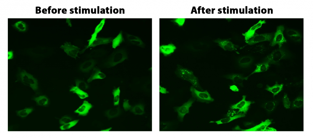
图1. HeLa细胞中β-arrestin的易位。 HeLa细胞用β-arrestin-GFP和加压素受体2(V2R)瞬时转染。 HeLa细胞在6孔板中培养,并融合至〜60%。用9 µL Transfectamine 5000转染等量的β-arrestin-GFP(1.5 µg)和V2R质粒(1.5 µg)。转染后约30小时,将细胞转移到96孔板中。转染后约48小时,将加压素(1 µM)加入细胞中以诱导β-arrestin-GFP易位。使用FITC通道在荧光显微镜下加压素处理之前和之后2小时拍摄图像。


参考文献

Screening cellular feature measurements for image-based assay development.
Authors: Logan, David J and Carpenter, Anne E
Journal: Journal of biomolecular screening (2010): 840-6

Kappa opioid receptor screen with the Tango beta-arrestin recruitment technology and characterization of hits with second-messenger assays.
Authors: Doucette, Christopher and Vedvik, Kevin and Koepnick, Elizabeth and Bergsma, Aaron and Thomson, Brian and Turek-Etienne, Tammy C
Journal: Journal of biomolecular screening (2009): 381-94

Multiplexed assays by high-content imaging for assessment of GPCR activity.
Authors: Ross, D A and Lee, S and Reiser, V and Xue, J and Alves, K and Vaidya, S and Kreamer, A and Mull, R and Hudak, E and Hare, T and Detmers, P A and Lingham, R and Ferrer, M and Strulovici, B and Santini, F
Journal: Journal of biomolecular screening (2008): 449-55

Comparison of G-protein coupled receptor desensitization-related beta-arrestin redistribution using confocal and non-confocal imaging.
Authors: Haasen, Dorothea and Wolff, Michael and Valler, Martin J and Heilker, Ralf
Journal: Combinatorial chemistry & high throughput screening (2006): 37-47

High-content screening of known G protein-coupled receptors by arrestin translocation.
Authors: Hudson, Christine C and Oakley, Robert H and Sjaastad, Michael D and Loomis, Carson R
Journal: Methods in enzymology (2006): 63-78

High-throughput confocal microscopy for beta-arrestin-green fluorescent protein translocation G protein-coupled receptor assays using the Evotec Opera.
Authors: Garippa, Ralph J and Hoffman, Ann F and Gradl, Gabriele and Kirsch, Achim
Journal: Methods in enzymology (2006): 99-120

The ligand-independent translocation assay: an enabling technology for screening orphan G protein-coupled receptors by arrestin recruitment.
Authors: Oakley, Robert H and Hudson, Christine C and Sjaastad, Michael D and Loomis, Carson R
Journal: Methods in enzymology (2006): 50-63

Development and validation of algorithms for measuring G-protein coupled receptor activation in cells using the LSC-based imaging cytometer platform.
Authors: Ozawa, Kazuo and Hudson, Christine C and Wille, Kirsten R and Karaki, Sachiko and Oakley, Robert H
Journal: Cytometry. Part A : the journal of the International Society for Analytical Cytology (2005): 69-76

Quantitative cell-based high-content screening for vasopressin receptor agonists using transfluor technology.
Authors: Ghosh, Richik N and DeBiasio, Richard and Hudson, Christine C and Ramer, Everett R and Cowan, Conrad L and Oakley, Robert H
Journal: Journal of biomolecular screening (2005): 476-84

The cellular distribution of fluorescently labeled arrestins provides a robust, sensitive, and universal assay for screening G protein-coupled receptors.
Authors: Oakley, Robert H and Hudson, Christine C and Cruickshank, Rachael D and Meyers, Diane M and Payne, Richard E and Rhem, Shay M and Loomis, Carson R
Journal: Assay and drug development technologies (2002): 21-30

说明书
Cell Meter Beta-Arrestin蛋白质易位GPCR信号检测试剂盒.pdf

Cell Meter Beta-Arrestin蛋白质易位GPCR信号检测试剂盒 货号36391-AAT Bioquest荧光染料

上海金畔生物科技有限公司代理AAT Bioquest荧光染料全线产品,欢迎访问AAT Bioquest荧光染料官网了解更多信息。

Cell Meter Beta-Arrestin蛋白质易位GPCR信号检测试剂盒

Cell Meter Beta-Arrestin蛋白质易位GPCR信号检测试剂盒

Cell Meter Beta-Arrestin蛋白质易位GPCR信号检测试剂盒    货号36391 货号 36391 存储条件 在零下15度以下保存, 避免光照
规格 200 Tests 价格 11628
Ex (nm) Em (nm)
分子量 溶剂
产品详细介绍

简要概述

所有G蛋白偶联受体(GPCR)在通过配体结合激活后,都会通过一条共同途径迅速发生脱敏作用。 β-arrestin(一种细胞质蛋白)与激活的受体的结合使GPCR信号失活,并启动了受体向细胞内的易位,在此细胞中配体被去除,受体被循环回到细胞膜。通过将荧光标记(例如GFP)附着到β-arrestin,可以检测受体arrestin复合物的位置。由于脱敏仅发生在激活的受体上,因此检测β-arrestin的转运和随后的受体再循环提供了检测GPCR靶标激活的可靠方法。 Cell Meter Beta-Arrestin蛋白易位GPCR信号试剂盒提供了功能强大的功能分析,可通过荧光成像筛选目标化合物针对已知或孤立GPCR靶标的活性。靶向GPCR的激活诱导荧光向细胞膜和内吞囊泡的移位。


适用仪器


荧光显微镜  
激发: FITC滤波片
发射: FITC滤波片
推荐孔板: 黑色透明底板

产品说明书

样品实验方案

概述

准备细胞进行转染
准备Transfectamine 5000-DNA混合物
将Transfectamine 5000-DNA混合物添加到细胞培养物中,并孵育过夜
转染后24-30小时将转染的细胞转移到96孔板中,并将培养物孵育过夜
在荧光显微镜下分析由GPCR激活引起的转运


细胞准备

细胞密度在转染时它们将达到约60-70%的融合度。
转染前用新鲜的生长培养基替换。
注意:例如,对于6孔板,每孔替换为2 mL培养基,对于10 cm板,则替换为6 mL培养基。


储备溶液配制

除非另有说明,否则所有未使用的储备溶液应分为一次性使用的等分试样,并在制备后储存在-20°C下。避免重复冻融循环。

β-arrestin-GFPDNA储备溶液
向小瓶β-arrestin-GFPDNA(组分A)中加入10 µL ddH2O,充分混合至终浓度为1 µg / µL。


操作步骤

1.将3 µg DNA(例如1.5 µg Beta-arrestin-GFP DNA储备液和1.5 µg您准备的GPCR DNA)与200 µL无血清培养基混合。
2.向混合物中加入9 µL Transfectamine 5000(组分B)。
3.充分混合并在室温下孵育20分钟。
注意:Transfectamine 5000和DNA的比例需要针对不同的细胞系进行优化,通常,在我们的测试中,Transfectamine 5000转染试剂(µL)与DNA(µg)的比例应为3-5 µL:1ug。

表1. 6孔板和10 cm板的样本表

  6孔板(每孔) 10cm板
新鲜培养基 2 mL 6 mL
质粒 3 µg 10 µg
无血清培养基 200 µL 600 µL
Transfectamine 5000转染试剂 ~9 µL ~30 µL


转染和转运方案
1.将Transfectamine 5000 -DNA混合物添加到培养板中,并孵育过夜。
注意事项:最早在转染后16小时即可检测到重组蛋白。转染后72〜96小时可观察到最大表达水平。
2.转染后24-30小时将转染的细胞转移到96孔板中,并孵育过夜。
使用FITC滤光片(Ex / Em = 488/530 nm)在荧光显微镜下检测受体激活诱导的β-arrestin易位。


图示

Cell Meter Beta-Arrestin蛋白质易位GPCR信号检测试剂盒    货号36391

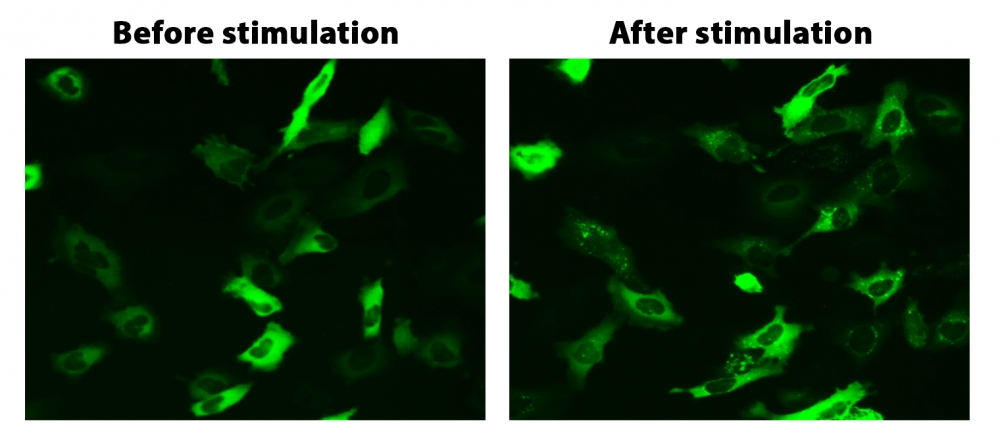
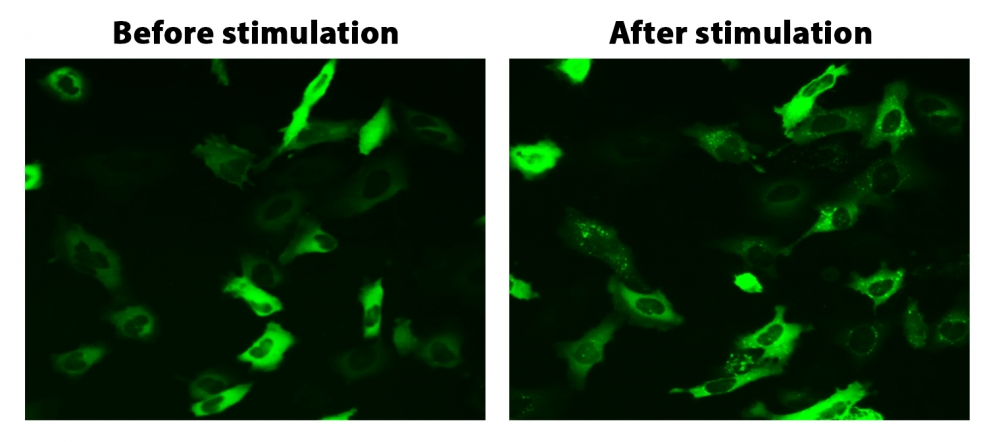
图1. HeLa细胞中β-arrestin的易位。 HeLa细胞用β-arrestin-GFP和加压素受体2(V2R)瞬时转染。 HeLa细胞在6孔板中培养,并融合至〜60%。用9 µL Transfectamine 5000转染等量的β-arrestin-GFP(1.5 µg)和V2R质粒(1.5 µg)。转染后约30小时,将细胞转移到96孔板中。转染后约48小时,将加压素(1 µM)加入细胞中以诱导β-arrestin-GFP易位。使用FITC通道在荧光显微镜下加压素处理之前和之后2小时拍摄图像。


参考文献

Screening cellular feature measurements for image-based assay development.
Authors: Logan, David J and Carpenter, Anne E
Journal: Journal of biomolecular screening (2010): 840-6

Kappa opioid receptor screen with the Tango beta-arrestin recruitment technology and characterization of hits with second-messenger assays.
Authors: Doucette, Christopher and Vedvik, Kevin and Koepnick, Elizabeth and Bergsma, Aaron and Thomson, Brian and Turek-Etienne, Tammy C
Journal: Journal of biomolecular screening (2009): 381-94

Multiplexed assays by high-content imaging for assessment of GPCR activity.
Authors: Ross, D A and Lee, S and Reiser, V and Xue, J and Alves, K and Vaidya, S and Kreamer, A and Mull, R and Hudak, E and Hare, T and Detmers, P A and Lingham, R and Ferrer, M and Strulovici, B and Santini, F
Journal: Journal of biomolecular screening (2008): 449-55

Comparison of G-protein coupled receptor desensitization-related beta-arrestin redistribution using confocal and non-confocal imaging.
Authors: Haasen, Dorothea and Wolff, Michael and Valler, Martin J and Heilker, Ralf
Journal: Combinatorial chemistry & high throughput screening (2006): 37-47

High-content screening of known G protein-coupled receptors by arrestin translocation.
Authors: Hudson, Christine C and Oakley, Robert H and Sjaastad, Michael D and Loomis, Carson R
Journal: Methods in enzymology (2006): 63-78

High-throughput confocal microscopy for beta-arrestin-green fluorescent protein translocation G protein-coupled receptor assays using the Evotec Opera.
Authors: Garippa, Ralph J and Hoffman, Ann F and Gradl, Gabriele and Kirsch, Achim
Journal: Methods in enzymology (2006): 99-120

The ligand-independent translocation assay: an enabling technology for screening orphan G protein-coupled receptors by arrestin recruitment.
Authors: Oakley, Robert H and Hudson, Christine C and Sjaastad, Michael D and Loomis, Carson R
Journal: Methods in enzymology (2006): 50-63

Development and validation of algorithms for measuring G-protein coupled receptor activation in cells using the LSC-based imaging cytometer platform.
Authors: Ozawa, Kazuo and Hudson, Christine C and Wille, Kirsten R and Karaki, Sachiko and Oakley, Robert H
Journal: Cytometry. Part A : the journal of the International Society for Analytical Cytology (2005): 69-76

Quantitative cell-based high-content screening for vasopressin receptor agonists using transfluor technology.
Authors: Ghosh, Richik N and DeBiasio, Richard and Hudson, Christine C and Ramer, Everett R and Cowan, Conrad L and Oakley, Robert H
Journal: Journal of biomolecular screening (2005): 476-84

The cellular distribution of fluorescently labeled arrestins provides a robust, sensitive, and universal assay for screening G protein-coupled receptors.
Authors: Oakley, Robert H and Hudson, Christine C and Cruickshank, Rachael D and Meyers, Diane M and Payne, Richard E and Rhem, Shay M and Loomis, Carson R
Journal: Assay and drug development technologies (2002): 21-30

说明书
Cell Meter Beta-Arrestin蛋白质易位GPCR信号检测试剂盒.pdf

FAM-xtra 亚磷酰胺 货号6037-AAT Bioquest荧光染料

上海金畔生物科技有限公司代理AAT Bioquest荧光染料全线产品,欢迎访问AAT Bioquest荧光染料官网了解更多信息。

FAM-xtra 亚磷酰胺

FAM-xtra 亚磷酰胺

FAM-xtra 亚磷酰胺    货号6037 货号 6037 存储条件 在零下15度以下保存, 避免光照
规格 50 umoles 价格 1164
Ex (nm) 493 Em (nm) 517
分子量 879.94 溶剂 MeCN
产品详细介绍

简要概述

产品基本信息

货号:6037

产品名称:FAM-xtra 亚磷酰胺

规格:50 umoles

储存条件:-15℃避光防潮

保质期:12个月


产品物理化学光谱特性

分子量:879.94

溶剂:MeCN


产品介绍

6-FAM亚磷酰胺可能是流行的荧光构建基块,已广泛用于通过荧光素荧光团有效标记5’端的寡核苷酸。标准裂解和氢氧化铵脱保护用于释放荧光素标记的寡核苷酸。尽管6-FAM已普遍用于开发各种qPCR探针,但其荧光受到两个作用限制:1.高度依赖pH值;2.光漂白迅速。目前我们已开发出FAM-xtra 亚磷酰胺解决了这两个局限性,同时完整保留了6-FAM亚磷酰胺的所有优点。 FAM-xtra亚磷酰胺可在相同操作条件下与6-FAM亚磷酰胺完全一样​​使用,而无需进行任何更改。由FAM-xtra亚磷酰胺制成的寡核苷酸具有许多优点:1.FAM-xtra标记的寡核苷酸具有与6-FAM标记的寡核苷酸相同的光谱。 2.FAM-xtra标记的寡核苷酸比相应的FAM-标记的寡核苷酸明亮得多。 3.FAM-xtra标记的寡核苷酸比相应的FAM-标记的寡核苷酸具有更高的光稳定性。4.与相应的FAM标记的寡核苷酸相比,FAM-xtra标记的寡核苷酸的荧光对pH的敏感度低得多。在生理条件下,FAM-xtra标记的寡核苷酸的荧光基本上与pH无关,这使得FAM-xtra标记的寡核苷酸的qPCR探针更加稳定。

点击查看光谱


参考文献

Development of an Innovative Intradermal siRNA Delivery System Using a Combination of a Functional Stearylated Cytoplasm-Responsive Peptide and a Tight Junction-Opening Peptide.
Authors: Ibaraki, Hisako and Kanazawa, Takanori and Takashima, Yuuki and Okada, Hiroaki and Seta, Yasuo
Journal: Molecules (Basel, Switzerland) (2016)

Non-nucleoside phosphoramidites of xanthene dyes (FAM, JOE, and TAMRA) for oligonucleotide labeling.
Authors: Kvach, Maksim V and Tsybulsky, Dmitry A and Shmanai, Vadim V and Prokhorenko, Igor A and Stepanova, Irina A and Korshun, Vladimir A
Journal: Current protocols in nucleic acid chemistry (2013): Unit 4.55

Practical synthesis of isomerically pure 5- and 6-carboxytetramethylrhodamines, useful dyes for DNA probes.
Authors: Kvach, Maksim V and Stepanova, Irina A and Prokhorenko, Igor A and Stupak, Aleksander P and Bolibrukh, Dmitry A and Korshun, Vladimir A and Shmanai, Vadim V
Journal: Bioconjugate chemistry (2009): 1673-82

Synthesis and cellular uptake of fluorescently labeled multivalent hyaluronan disaccharide conjugates of oligonucleotide phosphorothioates.
Authors: Karskela, Marika and Virta, Pasi and Malinen, Melina and Urtti, Arto and Lönnberg, Harri
Journal: Bioconjugate chemistry (2008): 2549-58

Synthesis of HyBeacons and dual-labelled probes containing 2′-fluorescent groups for use in genetic analysis.
Authors: Dobson, Neil and McDowell, David G and French, David J and Brown, Lynda J and Mellor, John M and Brown, Tom
Journal: Chemical communications (Cambridge, England) (2003): 1234-5

Amplified fragment length polymorphism (AFLP) protocol for genotyping the malarial parasite Plasmodium falciparum.
Authors: Rubio, J M and Berzosa, P J and Benito, A
Journal: Parasitology (2001): 331-6

Recovery of an oligonucleotide using silver ions immobilized onto paramagnetic particles.
Authors: Ramírez-Vick, J E and García, A A and Lee, J
Journal: Preparative biochemistry & biotechnology (1998): 243-60

Fluorescent dye phosphoramidite labelling of oligonucleotides.
Authors: Theisen, P and McCollum, C and Andrus, A
Journal: Nucleic acids symposium series (1992): 99-100

说明书
FAM-xtra 亚磷酰胺.pdf

FAM-xtra 亚磷酰胺 货号6038-AAT Bioquest荧光染料

上海金畔生物科技有限公司代理AAT Bioquest荧光染料全线产品,欢迎访问AAT Bioquest荧光染料官网了解更多信息。

FAM-xtra 亚磷酰胺

FAM-xtra 亚磷酰胺

FAM-xtra 亚磷酰胺    货号6038 货号 6038 存储条件 在零下15度以下保存, 避免光照
规格 100 umoles 价格 1776
Ex (nm) 493 Em (nm) 517
分子量 879.94 溶剂 MeCN
产品详细介绍

简要概述

产品基本信息

货号:6038

产品名称:FAM-xtra 亚磷酰胺

规格:100 umoles

储存条件:-15℃避光防潮

保质期:12个月


产品物理化学光谱特性

分子量:879.94

溶剂:MeCN


产品介绍

6-FAM亚磷酰胺可能是流行的荧光构建基块,已广泛用于通过荧光素荧光团有效标记5’端的寡核苷酸。标准裂解和氢氧化铵脱保护用于释放荧光素标记的寡核苷酸。尽管6-FAM已普遍用于开发各种qPCR探针,但其荧光受到两个作用限制:1.高度依赖pH值;2.光漂白迅速。目前我们已开发出FAM-xtra 亚磷酰胺解决了这两个局限性,同时完整保留了6-FAM亚磷酰胺的所有优点。 FAM-xtra亚磷酰胺可在相同操作条件下与6-FAM亚磷酰胺完全一样​​使用,而无需进行任何更改。由FAM-xtra亚磷酰胺制成的寡核苷酸具有许多优点:1.FAM-xtra标记的寡核苷酸具有与6-FAM标记的寡核苷酸相同的光谱。 2.FAM-xtra标记的寡核苷酸比相应的FAM-标记的寡核苷酸明亮得多。 3.FAM-xtra标记的寡核苷酸比相应的FAM-标记的寡核苷酸具有更高的光稳定性。4.与相应的FAM标记的寡核苷酸相比,FAM-xtra标记的寡核苷酸的荧光对pH的敏感度低得多。在生理条件下,FAM-xtra标记的寡核苷酸的荧光基本上与pH无关,这使得FAM-xtra标记的寡核苷酸的qPCR探针更加稳定。

点击查看光谱


参考文献

Development of an Innovative Intradermal siRNA Delivery System Using a Combination of a Functional Stearylated Cytoplasm-Responsive Peptide and a Tight Junction-Opening Peptide.
Authors: Ibaraki, Hisako and Kanazawa, Takanori and Takashima, Yuuki and Okada, Hiroaki and Seta, Yasuo
Journal: Molecules (Basel, Switzerland) (2016)

Non-nucleoside phosphoramidites of xanthene dyes (FAM, JOE, and TAMRA) for oligonucleotide labeling.
Authors: Kvach, Maksim V and Tsybulsky, Dmitry A and Shmanai, Vadim V and Prokhorenko, Igor A and Stepanova, Irina A and Korshun, Vladimir A
Journal: Current protocols in nucleic acid chemistry (2013): Unit 4.55

Practical synthesis of isomerically pure 5- and 6-carboxytetramethylrhodamines, useful dyes for DNA probes.
Authors: Kvach, Maksim V and Stepanova, Irina A and Prokhorenko, Igor A and Stupak, Aleksander P and Bolibrukh, Dmitry A and Korshun, Vladimir A and Shmanai, Vadim V
Journal: Bioconjugate chemistry (2009): 1673-82

Synthesis and cellular uptake of fluorescently labeled multivalent hyaluronan disaccharide conjugates of oligonucleotide phosphorothioates.
Authors: Karskela, Marika and Virta, Pasi and Malinen, Melina and Urtti, Arto and Lönnberg, Harri
Journal: Bioconjugate chemistry (2008): 2549-58

Synthesis of HyBeacons and dual-labelled probes containing 2′-fluorescent groups for use in genetic analysis.
Authors: Dobson, Neil and McDowell, David G and French, David J and Brown, Lynda J and Mellor, John M and Brown, Tom
Journal: Chemical communications (Cambridge, England) (2003): 1234-5

Amplified fragment length polymorphism (AFLP) protocol for genotyping the malarial parasite Plasmodium falciparum.
Authors: Rubio, J M and Berzosa, P J and Benito, A
Journal: Parasitology (2001): 331-6

Recovery of an oligonucleotide using silver ions immobilized onto paramagnetic particles.
Authors: Ramírez-Vick, J E and García, A A and Lee, J
Journal: Preparative biochemistry & biotechnology (1998): 243-60

Fluorescent dye phosphoramidite labelling of oligonucleotides.
Authors: Theisen, P and McCollum, C and Andrus, A
Journal: Nucleic acids symposium series (1992): 99-100

说明书
FAM-xtra 亚磷酰胺.pdf

FAM-xtra 亚磷酰胺 货号6039-AAT Bioquest荧光染料

上海金畔生物科技有限公司代理AAT Bioquest荧光染料全线产品,欢迎访问AAT Bioquest荧光染料官网了解更多信息。

FAM-xtra 亚磷酰胺

FAM-xtra 亚磷酰胺

FAM-xtra 亚磷酰胺    货号6039 货号 6039 存储条件 在零下15度以下保存, 避免光照
规格 10×100 umoles 价格 17748
Ex (nm) 493 Em (nm) 517
分子量 879.94 溶剂 MeCN
产品详细介绍

简要概述

产品基本信息

货号:6039

产品名称:FAM-xtra 亚磷酰胺

规格:10×100 umoles

储存条件:-15℃避光防潮

保质期:12个月


产品物理化学光谱特性

分子量:879.94

溶剂:MeCN


产品介绍

6-FAM亚磷酰胺可能是流行的荧光构建基块,已广泛用于通过荧光素荧光团有效标记5’端的寡核苷酸。标准裂解和氢氧化铵脱保护用于释放荧光素标记的寡核苷酸。尽管6-FAM已普遍用于开发各种qPCR探针,但其荧光受到两个作用限制:1.高度依赖pH值;2.光漂白迅速。目前我们已开发出FAM-xtra 亚磷酰胺解决了这两个局限性,同时完整保留了6-FAM亚磷酰胺的所有优点。 FAM-xtra亚磷酰胺可在相同操作条件下与6-FAM亚磷酰胺完全一样​​使用,而无需进行任何更改。由FAM-xtra亚磷酰胺制成的寡核苷酸具有许多优点:1.FAM-xtra标记的寡核苷酸具有与6-FAM标记的寡核苷酸相同的光谱。 2.FAM-xtra标记的寡核苷酸比相应的FAM-标记的寡核苷酸明亮得多。 3.FAM-xtra标记的寡核苷酸比相应的FAM-标记的寡核苷酸具有更高的光稳定性。4.与相应的FAM标记的寡核苷酸相比,FAM-xtra标记的寡核苷酸的荧光对pH的敏感度低得多。在生理条件下,FAM-xtra标记的寡核苷酸的荧光基本上与pH无关,这使得FAM-xtra标记的寡核苷酸的qPCR探针更加稳定。

点击查看光谱


参考文献

Development of an Innovative Intradermal siRNA Delivery System Using a Combination of a Functional Stearylated Cytoplasm-Responsive Peptide and a Tight Junction-Opening Peptide.
Authors: Ibaraki, Hisako and Kanazawa, Takanori and Takashima, Yuuki and Okada, Hiroaki and Seta, Yasuo
Journal: Molecules (Basel, Switzerland) (2016)

Non-nucleoside phosphoramidites of xanthene dyes (FAM, JOE, and TAMRA) for oligonucleotide labeling.
Authors: Kvach, Maksim V and Tsybulsky, Dmitry A and Shmanai, Vadim V and Prokhorenko, Igor A and Stepanova, Irina A and Korshun, Vladimir A
Journal: Current protocols in nucleic acid chemistry (2013): Unit 4.55

Practical synthesis of isomerically pure 5- and 6-carboxytetramethylrhodamines, useful dyes for DNA probes.
Authors: Kvach, Maksim V and Stepanova, Irina A and Prokhorenko, Igor A and Stupak, Aleksander P and Bolibrukh, Dmitry A and Korshun, Vladimir A and Shmanai, Vadim V
Journal: Bioconjugate chemistry (2009): 1673-82

Synthesis and cellular uptake of fluorescently labeled multivalent hyaluronan disaccharide conjugates of oligonucleotide phosphorothioates.
Authors: Karskela, Marika and Virta, Pasi and Malinen, Melina and Urtti, Arto and Lönnberg, Harri
Journal: Bioconjugate chemistry (2008): 2549-58

Synthesis of HyBeacons and dual-labelled probes containing 2′-fluorescent groups for use in genetic analysis.
Authors: Dobson, Neil and McDowell, David G and French, David J and Brown, Lynda J and Mellor, John M and Brown, Tom
Journal: Chemical communications (Cambridge, England) (2003): 1234-5

Amplified fragment length polymorphism (AFLP) protocol for genotyping the malarial parasite Plasmodium falciparum.
Authors: Rubio, J M and Berzosa, P J and Benito, A
Journal: Parasitology (2001): 331-6

Recovery of an oligonucleotide using silver ions immobilized onto paramagnetic particles.
Authors: Ramírez-Vick, J E and García, A A and Lee, J
Journal: Preparative biochemistry & biotechnology (1998): 243-60

Fluorescent dye phosphoramidite labelling of oligonucleotides.
Authors: Theisen, P and McCollum, C and Andrus, A
Journal: Nucleic acids symposium series (1992): 99-100

说明书
FAM-xtra 亚磷酰胺.pdf

ReadiView 绿色/红色 细胞活性染料 货号22763-AAT Bioquest荧光染料

上海金畔生物科技有限公司代理AAT Bioquest荧光染料全线产品,欢迎访问AAT Bioquest荧光染料官网了解更多信息。

ReadiView 绿色/红色 细胞活性染料

ReadiView 绿色/红色 细胞活性染料

ReadiView 绿色/红色 细胞活性染料    货号22763 货号 22763 存储条件 在零下15度以下保存, 避免光照
规格 1000 Tests 价格 6060
Ex (nm) Em (nm)
分子量 溶剂 Water
产品详细介绍

简要概述

ReadiView 绿色/红色活性染料是一种耐用的细胞染色剂,经过优化,可用于对Countess II FL或Countess 3 FL仪器中的有核细胞进行染色。 它可以方便地与传统的FITC或GFP滤光片组配合使用,例如EVOS GFP荧光灯适用于激发和检测绿色染料,而RFP或Texas Red(TxR)荧光灯适用于细胞红染色。 绿色表示活细胞,而红色表示死细胞。 ReadiView染料可对混合群体中的有核细胞进行特定染色,从而降低了复杂性,并在与适当的EVOS光学立方体匹配时提高了计数与样品间的可重复性。

产品说明书

样本实验方案

将10 µL ReadyCount 活/死活力染色剂与10 µL细胞混合。
在室温下孵育2至5分钟。
将10 µL染色的细胞样品上样至Countess细胞计数仪的玻片中。
在Countess细胞计数仪上读取数据。


图示

ReadiView 绿色/红色 细胞活性染料    货号22763

图1.用ReadyView 绿色/红色细胞活性染料标记的Jurkat细胞的荧光图像。


参考文献

Drug combinations against Borrelia burgdorferi persisters in vitro: eradication achieved by using daptomycin, cefoperazone and doxycycline.
Authors: Feng, Jie and Auwaerter, Paul G and Zhang, Ying
Journal: PloS one (2015): e0117207

Growth inhibition of cultured marine phytoplankton by toxic algal-derived polyunsaturated aldehydes.
Authors: Ribalet, François and Berges, John A and Ianora, Adrianna and Casotti, Raffaella
Journal: Aquatic toxicology (Amsterdam, Netherlands) (2007): 219-27

An improved method for the selective detection of fungi in hospital waters by solid phase cytometry.
Authors: De Vos, Muriel M and Nelis, Hans J
Journal: Journal of microbiological methods (2006): 557-65

A membrane-immunofluorescent-viability staining technique for the detection of Salmonella spp. from fresh and processed meat samples.
Authors: Duffy, G and Kilbride, B and Sheridan, J J and Blair, I S and McDowell, D A
Journal: Journal of applied microbiology (2000): 587-94

Staining with fluorescein diacetate correlates with hepatocyte function.
Authors: Nyberg, S L and Shatford, R A and Payne, W D and Hu, W S and Cerra, F B
Journal: Biotechnic & histochemistry : official publication of the Biological Stain Commission (1993): 56-63

说明书
ReadiView 绿色/红色 细胞活性染料.pdf

6-ROXtra,酸 货号386-AAT Bioquest荧光染料

上海金畔生物科技有限公司代理AAT Bioquest荧光染料全线产品,欢迎访问AAT Bioquest荧光染料官网了解更多信息。

6-ROXtra,酸

6-ROXtra,酸

货号 386 存储条件 在零下15度以下保存, 避免光照
规格 25 mg 价格 3612
Ex (nm) 578 Em (nm) 595
分子量 1034.34 溶剂 DMSO
产品详细介绍

简要概述

产品基本信息

货号:386

产品名称:6-ROXtra,酸

规格:25mg

储存条件:-15℃避光防潮

保质期:12个月


产品物理化学光谱特性

分子量:730.77

溶剂:DMSO

激发波长(nm):578

发射波长(nm):595


产品介绍

ROX染料具有很强的荧光性,但它们非常不稳定,一些商业样品的保质期只有几个月。 与6-ROX化合物相比,我们的6-ROXtra 染料具有更高的稳定性,同时与生物底物(例如寡核苷酸)缀合时具有与6-ROX基本上相同的光谱特性。 此外,我们的6-ROXtra 染料比基本上不溶于水和水性缓冲液的6-ROX染料水溶性更高。 我们的初步研究表明,在大多数情况下,6-ROXtra衍生的寡核苷酸比相应的6-ROX寡核苷酸更容易纯化。 当与蛋白质缀合时,6-ROX的荧光会被严重淬灭,而6-ROXtra 仍会显示非常强的荧光。

点击查看光谱


参考文献

Aligned Expression of IFI16 and STING Genes in RRMS Patients’ Blood.
Authors: Helbi, Sobhan and Ravanbakhsh, Behnam and Karimi, Mohammad and Kooti, Wesam and Jivad, Nahid
Journal: Endocrine, metabolic & immune disorders drug targets (2020): 878-886

RT-qPCR Detection of Low-Copy HIV RNA with Yin-Yang Probes.
Authors: Kireev, Dmitry E and Farzan, Valentina M and Shipulin, German A and Korshun, Vladimir A and Zatsepin, Timofei S
Journal: Methods in molecular biology (Clifton, N.J.) (2020): 27-35

SNPs and transcriptional activity of genes of innate and adaptive immunity at the maternal-fetal interface in woman with preterm labour, associated with preterm premature rupture of membranes.
Authors: Lyubomirskaya, Ekaterina S and Kamyshnyi, Alexandr M and Krut, Yuriy Ya and Smiianov, Vladyslav A and Fedoniuk, Larisa Ya and Romanyuk, Lidiya B and Kravets, Natalya Ya and Mochulska, Oksana M
Journal: Wiadomosci lekarskie (Warsaw, Poland : 1960) (2020): 25-30

Selenoprotein P inhibits cell proliferation and ROX production in HCC cells.
Authors: Wang, Jianxin and Shen, Pei and Liao, Sha and Duan, Lian and Zhu, Dandan and Chen, Jinling and Chen, Liuting and Sun, Xiaolei and Duan, Yinong
Journal: PloS one (2020): e0236491

Development of a multiplex TaqMan qPCR assay for simultaneous detection and differentiation of four DNA and RNA viruses from clinical samples of sheep and goats.
Authors: Xu, Xingang and Yang, Feng and Zhang, Qi and Xu, Ying and Huang, Jiali and Fu, Mingzhe and Zhang, Weimin
Journal: Journal of virological methods (2019): 58-64

Novel homo Yin-Yang probes improve sensitivity in RT-qPCR detection of low copy HIV RNA.
Authors: Farzan, Valentina M and Kvach, Maksim V and Aparin, Ilya O and Kireev, Dmitry E and Prikazchikova, Tatiana A and Ustinov, Alexey V and Shmanai, Vadim V and Shipulin, German A and Korshun, Vladimir A and Zatsepin, Timofei S
Journal: Talanta (2019): 226-232

Expression of Plasma hsa-miR122 in HBV-Related Hepatocellular Carcinoma (HCC) in Vietnamese Patients.
Authors: Quoc, Nguyen Bao and Phuong, Nguyen Doan Nguyen and Ngan, Tang Kim and Linh, Nguyen Thi Minh and Cuong, Pham Hung and Chau, Nguyen Ngoc Bao
Journal: MicroRNA (Shariqah, United Arab Emirates) (2018): 92-99

Simultaneous detection and quantification of 19 diarrhea-related pathogens with a quantitative real-time PCR panel assay.
Authors: Wongboot, Warawan and Okada, Kazuhisa and Chantaroj, Siriporn and Kamjumphol, Watcharaporn and Hamada, Shigeyuki
Journal: Journal of microbiological methods (2018): 76-82

Overexpression of OsERF48 causes regulation of OsCML16, a calmodulin-like protein gene that enhances root growth and drought tolerance.
Authors: Jung, Harin and Chung, Pil Joong and Park, Su-Hyun and Redillas, Mark Christian Felipe Reveche and Kim, Youn Shic and Suh, Joo-Won and Kim, Ju-Kon
Journal: Plant biotechnology journal (2017): 1295-1308

[Short-term Effect of Roxithromycin on Abundance and Diversity of Ammonia-Oxidizing Microorganisms in Activated Sludge].
Authors: Gao, Jing-Feng and Sun, Li-Xin and Fan, Xiao-Yan and Pan, Kai-Ling and Li, Ding-Chang
Journal: Huan jing ke xue= Huanjing kexue (2017): 2961-2971

说明书
6-ROXtra,酸.pdf

zFluor 635琥珀酰亚胺酯 货号1501-AAT Bioquest荧光染料

上海金畔生物科技有限公司代理AAT Bioquest荧光染料全线产品,欢迎访问AAT Bioquest荧光染料官网了解更多信息。

zFluor 635琥珀酰亚胺酯

zFluor 635琥珀酰亚胺酯

货号 1501 存储条件 在零下15度以下保存, 避免光照
规格 1 mg 价格 3000
Ex (nm) 667 Em (nm) 692
分子量 1336.89 溶剂 DMSO
产品详细介绍

简要概述

产品基本信息

货号:1501

产品名称:zFluor 635琥珀酰亚胺酯

规格:1mg

储存条件:-15℃避光防潮

保质期:12个月


产品物理化学光谱特性

分子量:1336.89

溶剂:DMSO

激发波长(nm):667

发射波长(nm):692


产品介绍

与市场上其他同类波长的染料相比,我们的zFluor 系列染料经过开发可在给定波长下具有最高的光稳定性。 zFluor 635具有与流行的Cy5®(GE Healthcare)和AlexaFluor®647(ThermoFisher)相似的光谱特性。其非常高的热稳定性和光稳定性使其成为单分子检测应用和高分辨率显微镜(如PALM,dSTORM和STED)的绝佳选择。在相同的测试条件下,zFluor 635的臭氧稳定性比Cy5®和AlexaFluor®647高得多,这使其成为微孔板和其他基于生物研究应用的更好选择。此功能对于荧光原位杂交(FISH)极为有用。此外,zFluor 标记的寡核苷酸和肽段比AlexaFluor®647和Cy5®标记的寡核苷酸和肽段更明亮,更光稳定。同样,在我们的qPCR测试中,zFluor 635标记的寡核苷酸表现出比用Cy5®或AlexaFluor®647制备的相应缀合物更高的信号。

点击查看光谱

说明书
zFluor 635琥珀酰亚胺酯.pdf

生物素NTA CAS 176046-06-3 货号3008-AAT Bioquest荧光染料

上海金畔生物科技有限公司代理AAT Bioquest荧光染料全线产品,欢迎访问AAT Bioquest荧光染料官网了解更多信息。

生物素NTA CAS 176046-06-3

生物素NTA CAS 176046-06-3

生物素NTA CAS 176046-06-3    货号3008 货号 3008 存储条件 在零下15度以下保存, 避免光照
规格 5 mg 价格 1164
Ex (nm) Em (nm)
分子量 488.56 溶剂 DMSO
产品详细介绍

简要概述

产品基本信息

货号:3008

产品名称:生物素NTA

CAS:176046-06-3

规格:5mg

储存条件:-15℃避光防潮

保质期:12个月


产品物理化学光谱特性

分子量:488.56

溶剂:DMSO


产品介绍

生物素标记的次氮基三乙酸生物素NTA(BNTA)被广泛用于检测固定在硝酸纤维素膜上的组氨酸标签蛋白,检测限为〜0.1 pmol。 NTA生物素是一种双功能试剂,用于检测固定的组氨酸标签蛋白。 次氮基三乙酸用于在其六个配位点中的四处螯合Ni(II)离子,其余两个位点可用于结合组氨酸标签。 可以使用标准的酶联链霉亲和素方法(例如链霉亲和素-辣根过氧化物酶偶联物)检测NTA-polyHis-复合物。 可以从pH 4.8的组氨酸标签蛋白中除去生物素NTA,从而可以用另一种探针重新分析印迹。 与荧光抗生物素蛋白结合物结合使用时,该NTA生物素衍生物可用于检测含多组氨酸的生物分子,例如融合蛋白。

说明书
生物素NTA CAS 176046-06-3.pdf

DiPO -1 [相当于POPO®-1] * 1 mM DMSO溶液* CAS 169454-15-3 货号17582-AAT Bioquest荧光染料

上海金畔生物科技有限公司代理AAT Bioquest荧光染料全线产品,欢迎访问AAT Bioquest荧光染料官网了解更多信息。

DiPO -1 [相当于POPO®-1] * 1 mM DMSO溶液* CAS 169454-15-3

DiPO -1 [相当于POPO®-1] * 1 mM DMSO溶液* CAS 169454-15-3

DiPO -1 [相当于POPO®-1] * 1 mM DMSO溶液* CAS 169454-15-3    货号17582 货号 17582 存储条件 在零下15度以下保存, 避免光照
规格 100 ul 价格 2388
Ex (nm) 433 Em (nm) 457
分子量 1170.54 溶剂 Water
产品详细介绍

简要概述

产品基本信息

货号:17582

产品名称:DiPO -1

CAS:169454-15-3

规格:100 ul

储存条件:-15℃避光防潮

保质期:24个月


产品物理化学光谱特性

分子量:1170.54

溶剂:DMSO

激发波长(nm):433

发射波长(nm):457


产品介绍

DiPO -1是与POPO®-1相同的化合物(POPO®是ThermoFisher的商标)。 DiPO -1是一种基于对称性花菁染料二聚体的高亲和力核酸染料,在不存在与核酸结合后表现出最大激发/发射最大〜434/456 nm的核酸的情况下,该染料基本不发荧光。 POPO-1是一种蓝色发射染料,可以被405激光激发,并被450/50滤光片激发,它通常用于流式细胞仪和荧光显微镜。

点击查看光谱


参考文献

Determination of restriction enzyme activity when cutting DNA labeled with the TOTO dye family.
Authors: Maschmann, April and Kounovsky-Shafer, Kristy L
Journal: Nucleosides, nucleotides & nucleic acids (2017): 406-417

Myosin Va and kinesin II motor proteins are concentrated in ribosomal domains (periaxoplasmic ribosomal plaques) of myelinated axons.
Authors: Sotelo-Silveira, José R and Calliari, Aldo and Cárdenas, Magdalena and Koenig, Edward and Sotelo, José R
Journal: Journal of neurobiology (2004): 187-96

Analysis of dimeric cyanine-nucleic acid dyes by capillary zone electrophoresis in N,N-dimethylacetamide as non-aqueous organic solvent.
Authors: Muzikar, Jan and Rozing, Gerard and van de Goor, Tom and Eberwein, Christine and Kenndler, Ernst
Journal: Journal of chromatography. A (2002): 249-55

Efficient detection of single DNA fragments in flowing sample streams by two-photon fluorescence excitation.
Authors: Van Orden, A and Cai, H and Goodwin, P M and Keller, R A
Journal: Analytical chemistry (1999): 2108-16

DNA-binding and fluorescence properties of the DNA bis-intercalating purple oxazole dimer POPO-1.
Authors: Winter, S and Loeber, G
Journal: Journal of biomedical optics (1997): 125-30

Fluorescently Labeled Virus Probes Show that Natural Virus Populations Can Control the Structure of Marine Microbial Communities.
Authors: Hennes, K P and Suttle, C A and Chan, A M
Journal: Applied and environmental microbiology (1995): 3623-7

说明书
DiPO -1 [相当于POPO®-1] * 1 mM DMSO溶液* CAS 169454-15-3.pdf

iFluor 488-dUTP * 1 mM的Tris缓冲液(pH 7.5)* 货号17039-AAT Bioquest荧光染料

上海金畔生物科技有限公司代理AAT Bioquest荧光染料全线产品,欢迎访问AAT Bioquest荧光染料官网了解更多信息。

iFluor 488-dUTP * 1 mM的Tris缓冲液(pH 7.5)*

iFluor 488-dUTP * 1 mM的Tris缓冲液(pH 7.5)*

iFluor 488-dUTP * 1 mM的Tris缓冲液(pH 7.5)*    货号17039 货号 17039 存储条件 在零下15度以下保存, 避免光照
规格 25 nmoles 价格 1164
Ex (nm) 491 Em (nm) 516
分子量 1152.83 溶剂 Water
产品详细介绍

简要概述

产品基本信息

货号:17039

产品名称:iFluor 488-dUTP

规格:25 nmoles

储存条件:-15℃避光防潮

保质期:24个月


产品物理化学光谱特性

分子量:1152.83

溶剂:水

激发波长(nm):491

发射波长(nm):516


产品介绍

染料修饰的脱氧尿苷5′-三磷酸酯是通过常规酶掺入方法(例如反转录,缺口翻译,随机引物标记或PCR)生产染料标记DNA的最常用方法之一。 这种酶促荧光标记方法广泛用于FISH探针和基于微阵列的实验。 该iFluor 488-dUTP缀合物与SpectrumGreen 滤光片组合一起用作绿色荧光(SpectrumGreen 是Vysis的商标)。 与常用的绿色探针(例如Fluorescein-12-dUTP)相比,iFluor 488提供了更明亮的信号,具有很高的光稳定性,并且不受pH值的影响。

点击查看光谱


参考文献

Anti-proliferative effect of Fe(III) complexed with 1-(2-hydroxy-3-methoxybenzaldehyde)-4-aminosalicylhydrazone in HepG2 cells.
Authors: Fukushima, Takeshi and Taniguchi, Erina and Yamada, Hiroshi and Kato, Kiyomasa and Shimizu, Ayako and Nishiguchi, Yoshikazu and Onozato, Mayu and Ichiba, Hideaki and Azuma, Yutaro
Journal: Biometals : an international journal on the role of metal ions in biology, biochemistry, and medicine (2015): 669-77

Localisation of abundant and organ-specific genes expressed in Rosa hybrida leaves and flower buds by direct in situ RT-PCR.
Authors: Jedrzejuk, Agata and Mibus, Heiko and Serek, Margrethe
Journal: TheScientificWorldJournal (2012): 609597

SNP genotyping using microsphere-linked PNA and flow cytometric detection.
Authors: Rockenbauer, Eszter and Petersen, Kenneth and Vogel, Ulla and Bolund, Lars and Kølvraa, Steen and Nielsen, Kirsten Vang and Nexø, Bjørn A
Journal: Cytometry. Part A : the journal of the International Society for Analytical Cytology (2005): 80-6

[Comparative genomic hybridization analysis of nasopharyngeal carcinoma drug-resistant cell CNE2/DDP and its parent cell CNE2].
Authors: Jiang, Run-De and Hu, Liang and Guan, Xin-Yuan and Zhang, Li-Xin and Yue, Wen and Cen, Xin-Tang and Li, Chun-Hai
Journal: Ai zheng = Aizheng = Chinese journal of cancer (2004): 386-90

Flow cytometric detection of beta-D-glucuronidase gene in wild-type bacterial cells using in-situ PCR.
Authors: Sachidanandham, Ramaiah and Gin, Karina Yew-Hoong
Journal: Biotechnology and bioengineering (2003): 127-33

Lack of nuclear apoptosis in cardiomyocytes and increased endothelin-1 levels in a rat heart model of myocardial stunning.
Authors: Klainguti, M and Aigner, S and Kilo, J and Eppenberger, H M and Mandinova, A and Aebi, U and Schaub, M C and Shaw, S G and Lüscher, T F and Atar, D
Journal: Basic research in cardiology (2000): 308-15

A method to monitor DNA transfer during transfection.
Authors: Johnson, A L and Jurcisek, J A and Trask, O J and Au, J L
Journal: AAPS pharmSci (1999): E6

Pulsatile shear stress leads to DNA fragmentation in human SH-SY5Y neuroblastoma cell line.
Authors: Triyoso, D H and Good, T A
Journal: The Journal of physiology (1999): 355-65

Detection of TNF alpha and Fas ligand mRNA within synovial mononuclear cells by fluorescence in-cell labeling PCR (FICL-PCR).
Authors: Okubo, M and Brown, M P and Chiba, K and Kasukawa, R and Nishimaki, T
Journal: Molecular biology reports (1998): 217-24

Targeted nucleotide exchange in the alkaline phosphatase gene of HuH-7 cells mediated by a chimeric RNA/DNA oligonucleotide.
Authors: Kren, B T and Cole-Strauss, A and Kmiec, E B and Steer, C J
Journal: Hepatology (Baltimore, Md.) (1997): 1462-8

说明书
iFluor 488-dUTP * 1 mM的Tris缓冲液(pH 7.5)*.pdf

Cell Meter 活细胞ATP检测试剂盒 货号23015-AAT Bioquest荧光染料

上海金畔生物科技有限公司代理AAT Bioquest荧光染料全线产品,欢迎访问AAT Bioquest荧光染料官网了解更多信息。

Cell Meter 活细胞ATP检测试剂盒

Cell Meter 活细胞ATP检测试剂盒

Cell Meter 活细胞ATP检测试剂盒    货号23015 货号 23015 存储条件 在零下15度以下保存, 避免光照
规格 100 Tests 价格 4224
Ex (nm) Em (nm)
分子量 溶剂
产品详细介绍

简要概述

三磷酸腺苷(ATP)在细胞能量、代谢调节和细胞信号传导中起着基本作用。它被称为细胞内能量转移的“货币分子单位”,以驱动活细胞中的许多生物过程和化学合成。 ATP也是细胞通讯的信号分子,在DNA和RNA合成中起着重要作用。它位于发生细胞呼吸的线粒体中。 ATP水平可用于测量细胞增殖和细胞周期动态。 Cell Meter 活细胞ATP检测试剂盒使研究人员可以使用检测细胞ATP的渗透型红色荧光成像探针ATP Red 来检测活细胞中的ATP水平。 ATP Red 旨在检测活细胞线粒体中的ATP浓度。该探针与AMP,ADP,CMP,CDP,CTP,UMP,UDP,UTP,GMP,GDP或GTP的交叉反应性最小。


适用仪器


荧光酶标仪  
激发: Cy3/TRITC滤波片组
发射: Cy3/TRITC滤波片组
推荐孔板: 黑色透明孔板

产品说明书

实验方案

概述

在生长培养基中准备细胞
将细胞与ATP Red 工作溶液在37°C下孵育15-30分钟
去掉ATP Red 工作溶液
使用Cy3 / TRITC滤波片组的荧光显微镜进行检测


准备细胞

对于贴壁细胞
在96孔板的生长培养基中以10,000至40,000个细胞/孔/ 90μL平板培养细胞过夜,而对于384孔板则以2,500至10,000个细胞/孔/ 20μL平板培养过夜。

对于悬浮细胞
离心培养液中的细胞,然后将细胞沉淀以50,000-100,000细胞/孔/ 90 µL的浓度悬浮于96孔聚D赖氨酸平板中,或10,000-25,000细胞/孔/ 20 / L以获得384孔中的悬浮液 聚-D赖氨酸板。 实验前,将板以800 rpm的速度离心2分钟。
注意:应该对每个细胞系进行单独评估,以确定最佳细胞密度。


溶液配制

除非另有说明,否则所有未使用的储备溶液应分为一次性使用的等分试样,并在制备后储存在-20°C下。 避免重复冻融循环。

ATP Red 储备溶液配制(500X)
将50 µL DMSO(组分C)添加到小瓶ATP Red (组分A)中,制成500X储备液。
注意:50 µL的500X ATP Red 储备溶液足以容纳一个96孔板。 可以将未使用的ATP Red 储备溶液分装,并在≤-20°C下以较小的等分试样存储。 避光,并避免重复的冻融循环。


ATP Red 工作溶液配制
将5 µL 500X储备溶液(组分A)添加到1 mL细胞培养基中,并充分混合。
注意:ATP Red 探针与我们检测的大多数细胞系的细胞培养基兼容。 可以根据特定的细胞类型修改染色条件。


操作步骤

1.在生长培养基中准备细胞。
2.在细胞板中加入等体积的[100 µL /孔(96孔板)或25 µL /孔(384孔板)]。
注意:ATP Red 的最佳浓度因具体应用而异。
3.在避光的条件下,将细胞在37°C下孵育15-30分钟。
4.从每个孔中除去工作溶液。 用测定缓冲液(组分B)或您选择的缓冲液洗涤细胞两次。
5.使用带有Cy3 / TRITC滤光片组的荧光显微镜观察细胞中的荧光信号。


图示

Cell Meter 活细胞ATP检测试剂盒    货号23015

图1. HeLa细胞与ATP Red 和MitoLite Green FM共染色的荧光图像在96孔黑色透明底板。 HeLa细胞用ATP Red 染色15分钟,然后与100 nM MitoLite Green FM(Cat#22695)孵育30分钟。 成像前用测定缓冲液洗涤两次。 ATP Red 和MitoLite ATP Red 信号重叠良好。使用带有Cy3 / TRITC和FITC滤光片的荧光显微镜对细胞成像。


参考文献

Metabolic co-dependence of the oocyte and cumulus cells: essential role in determining oocyte developmental competence.
Authors: Richani, Dulama and Dunning, Kylie R and Thompson, Jeremy G and Gilchrist, Robert B
Journal: Human reproduction update (2021): 27-47

Mitochondrial C3a Receptor Activation in Oxidatively Stressed Epithelial Cells Reduces Mitochondrial Respiration and Metabolism.
Authors: Ishii, Masaaki and Beeson, Gyda and Beeson, Craig and Rohrer, Bärbel
Journal: Frontiers in immunology (2021): 628062

Cell-penetrating and mitochondrion-targeting molecules.
Authors: Appiah Kubi, George and Pei, Dehua
Journal: Methods in enzymology (2020): 311-328

Combinatorial roles of mitochondria and cGMP/PKG pathway in the generation of neuronal free Zn2+ under the presence of nitric oxide.
Authors: Yang, De-Ming and Huang, Chien-Chang and Chang, Yu-Fen
Journal: Journal of the Chinese Medical Association : JCMA (2020): 357-366

Dynamic regulation of subcellular mitochondrial position for localized metabolite levels.
Authors: Alshaabi, Haya and Heininger, Meara and Cunniff, Brian
Journal: Journal of biochemistry (2020): 109-117

Engineering a Reversible Fluorescent Probe for Real-Time Live-Cell Imaging and Quantification of Mitochondrial ATP.
Authors: Ren, Tian-Bing and Wen, Si-Yu and Wang, Lu and Lu, Peng and Xiong, Bin and Yuan, Lin and Zhang, Xiao-Bing
Journal: Analytical chemistry (2020): 4681-4688

Heat stress induces apoptosis through disruption of dynamic mitochondrial networks in dairy cow mammary epithelial cells.
Authors: Chen, Kun-Lin and Wang, Hui-Li and Jiang, Lin-Zheng and Qian, Yong and Yang, Cai-Xia and Chang, Wei-Wei and Zhong, Ji-Feng and Xing, Guang-Dong
Journal: In vitro cellular & developmental biology. Animal (2020): 322-331

Intracellular ATP levels influence cell fates in Dictyostelium discoideum differentiation.
Authors: Hiraoka, Haruka and Nakano, Tadashi and Kuwana, Satoshi and Fukuzawa, Masashi and Hirano, Yasuhiro and Ueda, Masahiro and Haraguchi, Tokuko and Hiraoka, Yasushi
Journal: Genes to cells : devoted to molecular & cellular mechanisms (2020): 312-326

Measurement of ATP concentrations in mitochondria of living cells using luminescence and fluorescence approaches.
Authors: Morciano, Giampaolo and Imamura, Hiromi and Patergnani, Simone and Pedriali, Gaia and Giorgi, Carlotta and Pinton, Paolo
Journal: Methods in cell biology (2020): 199-219

Mitochondria transfer enhances proliferation, migration, and osteogenic differentiation of bone marrow mesenchymal stem cell and promotes bone defect healing.
Authors: Guo, Yusi and Chi, Xiaopei and Wang, Yifan and Heng, Boon Chin and Wei, Yan and Zhang, Xuehui and Zhao, Han and Yin, Ying and Deng, Xuliang
Journal: Stem cell research & therapy (2020): 245

说明书
Cell Meter 活细胞ATP检测试剂盒.pdf

GTTR[Texas Red 庆大霉素缀合物] 货号24300-AAT Bioquest荧光染料

上海金畔生物科技有限公司代理AAT Bioquest荧光染料全线产品,欢迎访问AAT Bioquest荧光染料官网了解更多信息。

GTTR[Texas Red 庆大霉素缀合物]

GTTR[Texas Red 庆大霉素缀合物]

GTTR[Texas Red 庆大霉素缀合物]    货号24300 货号 24300 存储条件 在零下15度以下保存, 避免光照
规格 100 ug 价格 2388
Ex (nm) 586 Em (nm) 603
分子量 ~1200 溶剂 DMSO
产品详细介绍

简要概述

产品基本信息

货号:24300

产品名称:GTTR[Texas Red 庆大霉素缀合物]

规格:100ug

储存条件:-15℃避光防潮

保质期:24个月


产品物理化学光谱特性

分子量:~1300

溶剂:DMSO

激发波长(nm):586

发射波长(nm):603


产品介绍

庆大霉素被广泛用于治疗结核病和革兰氏阴性感染,在新生儿重症监护中特别有用。带有荧光团标记的药物,例如庆大霉素-Texas Red(GTTR)缀合物,已被用于鉴定跨BLB进入毛细胞,特别是在耳蜗(负责听觉的感觉器官)中的药物运输途径。识别耳蜗内向耳细胞运输耳毒性药物所涉及的机制具有重要的基础和临床意义。Texas Red庆大霉素缀合物(GTTR)可通过荧光技术(例如荧光显微镜,荧光相关光谱法,单分子光谱法)研究庆大霉素的细胞吸收,动力学和分布。 TR荧光团可以方便地在500-600 nm范围内激发,最大发射集中在610 nm附近。

点击查看光谱


参考文献

Development of a green fluorescence protein (GFP)-based bioassay for detection of antibiotics and its application in milk.
Authors: Yazgan Karacaglar, Nazife Nur and Topcu, Ali and Dudak, Fahriye Ceyda and Boyaci, Ismail Hakki
Journal: Journal of food science (2020): 500-509

Micronomicin/tobramycin binding with DNA: fluorescence studies using of ethidium bromide as a probe and molecular docking analysis.
Authors: Bi, Shuyun and Zhou, Huifeng and Wu, Jun and Sun, Xiaoyue
Journal: Journal of biomolecular structure & dynamics (2019): 1464-1476

Label-free and nicking enzyme-assisted fluorescence signal amplification for RNase H determination based on a G-quadruplexe/thioflavin T complex.
Authors: Wu, Kefeng and Ma, Changbei and Deng, Zhiyi and Fang, Ning and Tang, Zhenwei and Zhu, Xingxing and Wang, Kemin
Journal: Talanta (2018): 142-147

Gentamicin differentially alters cellular metabolism of cochlear hair cells as revealed by NAD(P)H fluorescence lifetime imaging.
Authors: Zholudeva, Lyandysha V and Ward, Kristina G and Nichols, Michael G and Smith, Heather Jensen
Journal: Journal of biomedical optics (2015): 051032

Fluorescence microscopy methods for determining the viability of bacteria in association with mammalian cells.
Authors: Johnson, M Brittany and Criss, Alison K
Journal: Journal of visualized experiments : JoVE (2013)

Screening system of blocking agents of the receptor for advanced glycation endproducts in cells using fluorescence.
Authors: Jung, Dong Ho and Kim, Young Sook and Kim, Jin Sook
Journal: Biological & pharmaceutical bulletin (2012): 1826-30

[Determination of netilmicin in rat plasma by reversed-phase high performance liquid chromatography with fluorescence detection and pre-column derivatization].
Authors: Chang, Xiaojuan and Peng, Jingdong and Liu, Shaopu and Liu, Limin and Dai, Yongkuang
Journal: Se pu = Chinese journal of chromatography (2009): 794-8

[Fluorescence resonance energy transfer between gentamycin and water-soluble CdTe QDs].
Authors: Li, Jin-Gui and Zhu, Kui and Xu, Fei and Jiang, Hai-Yang and Ding, Shuang-Yang
Journal: Guang pu xue yu guang pu fen xi = Guang pu (2009): 3070-4

Determination of isepamicin in human plasma by HPLC with fluorescence detection after derivatization using 6-aminoquinolyl-N-hydroxysuccinimidyl-carbamate.
Authors: Hosokawa, Shin and Nakamura, Katsunori and Fujita, Yukiyoshi and Horiuchi, Ryuya and Yamamoto, Koujirou
Journal: Biological & pharmaceutical bulletin (2008): 1866-9

Flow injection fluorescence immunoassay for gentamicin using sol-gel-derived mesoporous biomaterial.
Authors: Yang, Huang-Hao and Zhu, Qing-Zhi and Qu, Hui-Ying and Chen, Xiao-Lan and Ding, Ma-Tai and Xu, Jin-Gou
Journal: Analytical biochemistry (2002): 71-6

说明书
GTTR[Texas Red 庆大霉素缀合物].pdf

GTTR[Texas Red 庆大霉素缀合物] 货号24301-AAT Bioquest荧光染料

上海金畔生物科技有限公司代理AAT Bioquest荧光染料全线产品,欢迎访问AAT Bioquest荧光染料官网了解更多信息。

GTTR[Texas Red 庆大霉素缀合物]

GTTR[Texas Red 庆大霉素缀合物]

GTTR[Texas Red 庆大霉素缀合物]    货号24301 货号 24301 存储条件 在零下15度以下保存, 避免光照
规格 1 mg 价格 11628
Ex (nm) 586 Em (nm) 603
分子量 ~1200 溶剂 DMSO
产品详细介绍

简要概述

产品基本信息

货号:24301

产品名称:GTTR[Texas Red 庆大霉素缀合物]

规格:1mg

储存条件:-15℃避光防潮

保质期:24个月


产品物理化学光谱特性

分子量:~1300

溶剂:DMSO

激发波长(nm):586

发射波长(nm):603


产品介绍

庆大霉素被广泛用于治疗结核病和革兰氏阴性感染,在新生儿重症监护中特别有用。带有荧光团标记的药物,例如庆大霉素-Texas Red(GTTR)缀合物,已被用于鉴定跨BLB进入毛细胞,特别是在耳蜗(负责听觉的感觉器官)中的药物运输途径。识别耳蜗内向耳细胞运输耳毒性药物所涉及的机制具有重要的基础和临床意义。Texas Red庆大霉素缀合物(GTTR)可通过荧光技术(例如荧光显微镜,荧光相关光谱法,单分子光谱法)研究庆大霉素的细胞吸收,动力学和分布。 TR荧光团可以方便地在500-600 nm范围内激发,最大发射集中在610 nm附近。

点击查看光谱


参考文献

Development of a green fluorescence protein (GFP)-based bioassay for detection of antibiotics and its application in milk.
Authors: Yazgan Karacaglar, Nazife Nur and Topcu, Ali and Dudak, Fahriye Ceyda and Boyaci, Ismail Hakki
Journal: Journal of food science (2020): 500-509

Micronomicin/tobramycin binding with DNA: fluorescence studies using of ethidium bromide as a probe and molecular docking analysis.
Authors: Bi, Shuyun and Zhou, Huifeng and Wu, Jun and Sun, Xiaoyue
Journal: Journal of biomolecular structure & dynamics (2019): 1464-1476

Label-free and nicking enzyme-assisted fluorescence signal amplification for RNase H determination based on a G-quadruplexe/thioflavin T complex.
Authors: Wu, Kefeng and Ma, Changbei and Deng, Zhiyi and Fang, Ning and Tang, Zhenwei and Zhu, Xingxing and Wang, Kemin
Journal: Talanta (2018): 142-147

Gentamicin differentially alters cellular metabolism of cochlear hair cells as revealed by NAD(P)H fluorescence lifetime imaging.
Authors: Zholudeva, Lyandysha V and Ward, Kristina G and Nichols, Michael G and Smith, Heather Jensen
Journal: Journal of biomedical optics (2015): 051032

Fluorescence microscopy methods for determining the viability of bacteria in association with mammalian cells.
Authors: Johnson, M Brittany and Criss, Alison K
Journal: Journal of visualized experiments : JoVE (2013)

Screening system of blocking agents of the receptor for advanced glycation endproducts in cells using fluorescence.
Authors: Jung, Dong Ho and Kim, Young Sook and Kim, Jin Sook
Journal: Biological & pharmaceutical bulletin (2012): 1826-30

[Determination of netilmicin in rat plasma by reversed-phase high performance liquid chromatography with fluorescence detection and pre-column derivatization].
Authors: Chang, Xiaojuan and Peng, Jingdong and Liu, Shaopu and Liu, Limin and Dai, Yongkuang
Journal: Se pu = Chinese journal of chromatography (2009): 794-8

[Fluorescence resonance energy transfer between gentamycin and water-soluble CdTe QDs].
Authors: Li, Jin-Gui and Zhu, Kui and Xu, Fei and Jiang, Hai-Yang and Ding, Shuang-Yang
Journal: Guang pu xue yu guang pu fen xi = Guang pu (2009): 3070-4

Determination of isepamicin in human plasma by HPLC with fluorescence detection after derivatization using 6-aminoquinolyl-N-hydroxysuccinimidyl-carbamate.
Authors: Hosokawa, Shin and Nakamura, Katsunori and Fujita, Yukiyoshi and Horiuchi, Ryuya and Yamamoto, Koujirou
Journal: Biological & pharmaceutical bulletin (2008): 1866-9

Flow injection fluorescence immunoassay for gentamicin using sol-gel-derived mesoporous biomaterial.
Authors: Yang, Huang-Hao and Zhu, Qing-Zhi and Qu, Hui-Ying and Chen, Xiao-Lan and Ding, Ma-Tai and Xu, Jin-Gou
Journal: Analytical biochemistry (2002): 71-6

说明书
GTTR[Texas Red 庆大霉素缀合物].pdf

Bucculite XdU细胞增殖检测试剂盒*紫色激光兼容**流式细胞术* 货号22321-AAT Bioquest荧光染料

上海金畔生物科技有限公司代理AAT Bioquest荧光染料全线产品,欢迎访问AAT Bioquest荧光染料官网了解更多信息。

Bucculite XdU细胞增殖检测试剂盒*紫色激光兼容**流式细胞术*

Bucculite XdU细胞增殖检测试剂盒*紫色激光兼容**流式细胞术*

Bucculite XdU细胞增殖检测试剂盒*紫色激光兼容**流式细胞术*    货号22321 货号 22321 存储条件 在零下15度以下保存, 避免光照
规格 100 Tests 价格 0
Ex (nm) Em (nm)
分子量 N/A 溶剂
产品详细介绍

简要概述

检测细胞增殖是评估细胞活力、细胞周期和遗传毒性的最可靠的方法之一。 该试剂盒采用的是检测细胞增殖的一种基本方法,它是在细胞生长的S期过程中,在存在胸腺嘧啶核苷的情况下检测DNA的合成。 Bucculite 流式细胞术XdU细胞增殖测定试剂盒使用XdU,该XdU在DNA合成过程中被掺入细胞DNA中。 固定细胞后,将掺入的XdU用mFluor Violet 450叠氮化物标记。 在Pacific Blue通道中观察mFluor 紫罗兰色450标记的DNA。 Bucculite 流式细胞术XdU细胞增殖检测试剂盒是抗BrdU抗体的检测和EdU点击化学检测的的完美替代品。 它的灵敏度非常高,可用于在单细胞水平上检测活性DNA的合成。


适用仪器


流式细胞仪  
激发: 405nm laser
发射: 450/40nm 滤波片组
通道: Pacific Blue通道

产品说明书

样品实验方案

概述

准备细胞
使用XdU工作溶液处理细胞3小时
固定细胞
透化细胞
用Bucculite 工作溶液处理细胞30分钟
在流式细胞仪中使用450/40 nm滤光片组观察荧光


溶液配制

1.储备溶液配制

1.1 XdU储备溶液(100X)
将XdU(组分A)溶于200 µL DMSO中制成XdU储备溶液。
注意:可以将剩余的XdU储备溶液以较小的等分试样在-20°C下存储。
注意:避免光线直射,并避免重复的冻融循环。

1.2mFluor 紫色450-叠氮化物储备溶液(200 X)
将mFluor 紫色450-叠氮化物(组分B)溶于100 µL DMSO中,制成mFluor 紫色450-叠氮化物储备溶液。
注意:可以将剩余的mFluor 紫色450叠氮化物储备溶液以较小的等分试样在-20°C下存储。
注意:避免光线直射,并避免重复的冻融循环。

1.3XdU反应添加剂储备溶液(20X)
将XdU反应添加剂(组分E)溶于1 mL去离子水中,制成XdU反应添加剂储备溶液。
注意:剩余的XdU反应添加剂储备溶液可以在-20°C下保存。


2.工作溶液配制

2.1XdU工作溶液配制
在1 mL细胞培养基中加入10 µL 100X XdU储备溶液,制成XdU工作溶液。
注意:1 mL的工作溶液足以进行5次检测。
注意:此100X浓度是使用优化的XdU浓度在HeLa和Jurkat细胞中测试的。 生长培养基、细胞密度、细胞类型变化和其他因素可能会影响标记。 我们建议检测一系列XdU浓度,以确定最佳浓度以及是否适合您的细胞等因素。

2.2Bucculite 工作溶液
按以下顺序混合并充分涡旋
①905 µL染色缓冲液(组分C)
②40 µL CuSO4(组分D)
③5 µL mFluor 紫色450叠氮化物储备溶液
④50 µL XdU反应添加剂储备溶液
注意:1 mL的Bucculite 工作溶液足以进行5次检测。


操作步骤

1.用XdU标记细胞
考虑到细胞在200 µL细胞培养基中,请向细胞中加入200 µL XdU工作溶液。
在适合细胞类型的最佳条件下,将细胞孵育3小时。 XdU在细胞中的时间可以直接检测细胞DNA的合成。 孵育时间取决于细胞生长速率。


2.细胞固定
孵育后,除去培养基,并用PBS洗涤细胞一次。
注意:清洗步骤后,如果使用贴壁细胞,请使用ReadiUse 细胞分离缓冲液(#60010)分离它们。
向每个试管中加入200 µL的固定缓冲液(含3.7%甲醛的PBS溶液,未提供),并在室温下孵育15-20分钟。
除去固定液,并用PBS清洗每个试管中的细胞两次。


3.细胞通透性处理
向每个试管中加入200 µL透化缓冲液(0.5%Triton®X-100的PBS溶液,未提供),并在室温下孵育10-20分钟。
除去通透缓冲液,并用PBS清洗每个试管中的细胞两次。


4.细胞染色
在试管中加入200 µL Bucculite 工作溶液。 在室温避光下,将Bucculite 工作溶液在室温下孵育细胞30分钟。
除去每个管中的Bucculite 工作溶液。
用PBS洗涤细胞两次,洗涤后加入200 µL PBS。
在流式细胞仪中使用450/40 nm滤光片观察细胞中的荧光信号。


图示

Bucculite XdU细胞增殖检测试剂盒*紫色激光兼容**流式细胞术*    货号22321

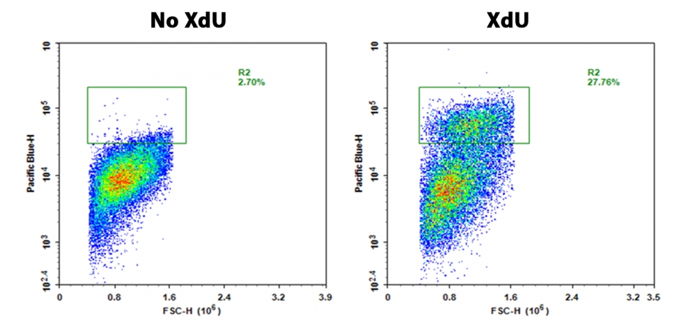
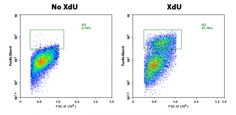
图1.用Buccutite XdU细胞增殖检测试剂盒(Cat#22321)检测到的S期Jurkat细胞的流式细胞仪中的反应。 将Jurkat细胞以50,000个细胞/孔/ 100μL的浓度接种在6孔黑色板上过夜。 按照说明书,将细胞在37℃下用XdU处理3小时,固定并透化。 然后将细胞在染色缓冲液中用mFluor 紫色450-叠氮化物染色30分钟,并用PBS洗涤3次。 使用Pacific Blue通道,通过NovoCyte流式细胞仪检测荧光响应。

说明书
Bucculite XdU细胞增殖检测试剂盒*紫色激光兼容**流式细胞术*.pdf

Bucculite XdU细胞增殖检测试剂盒*蓝色激光兼容**流式细胞术* 货号22323-AAT Bioquest荧光染料

上海金畔生物科技有限公司代理AAT Bioquest荧光染料全线产品,欢迎访问AAT Bioquest荧光染料官网了解更多信息。

Bucculite XdU细胞增殖检测试剂盒*蓝色激光兼容**流式细胞术*

Bucculite XdU细胞增殖检测试剂盒*蓝色激光兼容**流式细胞术*

Bucculite XdU细胞增殖检测试剂盒*蓝色激光兼容**流式细胞术*    货号22323 货号 22323 存储条件 在零下15度以下保存, 避免光照
规格 100 Tests 价格 6060
Ex (nm) Em (nm)
分子量 溶剂
产品详细介绍

简要概述

检测细胞增殖是评估细胞活力、细胞周期和遗传毒性的最可靠的方法之一。 该试剂盒采用的是检测细胞增殖的一种基本方法,它是在细胞生长的S期过程中,在存在胸腺嘧啶核苷的情况下检测DNA的合成。 Bucculite 流式细胞术XdU细胞增殖测定试剂盒使用XdU,该XdU在DNA合成过程中被掺入细胞DNA中。 固定细胞后,将掺入的XdU用iFluor 488叠氮化物标记。 在FITC通道中观察iFluor 488标记的DNA。 Bucculite 流式细胞术XdU细胞增殖检测试剂盒是抗BrdU抗体的检测和EdU点击化学检测的的完美替代品。 它的灵敏度非常高,可用于在单细胞水平上检测活性DNA的合成。


适用仪器


流式细胞仪  
激发: 488nm laser
发射: 530/30nm 滤波片组
通道: FITC通道

产品说明书

样品实验方案

概述

准备细胞
使用XdU工作溶液处理细胞3小时
固定细胞
透化细胞
用Bucculite 工作溶液处理细胞30分钟
在流式细胞仪中使用530/30 nm滤光片组观察荧光


溶液配制

1.储备溶液配制

1.1 XdU储备溶液(100X)
将XdU(组分A)溶于200 µL DMSO中制成XdU储备溶液。
注意:可以将剩余的XdU储备溶液以较小的等分试样在-20°C下存储。
注意:避免光线直射,并避免重复的冻融循环。

1.2iFluor 488叠氮化物储备溶液(200 X)
将iFluor 488叠氮化物(组分B)溶于100 µL DMSO中,制成iFluor 488叠氮化物储备溶液。
注意:可以将剩余的iFluor 488叠氮化物储备溶液以较小的等分试样在-20°C下存储。
注意:避免光线直射,并避免重复的冻融循环。

1.3XdU反应添加剂储备溶液(20X)
将XdU反应添加剂(组分E)溶于1 mL去离子水中,制成XdU反应添加剂储备溶液。
注意:剩余的XdU反应添加剂储备溶液可以在-20°C下保存。


2.工作溶液配制

2.1XdU工作溶液配制
在1 mL细胞培养基中加入10 µL 100X XdU储备溶液,制成XdU工作溶液。
注意:1 mL的工作溶液足以进行5次检测。
注意:此100X浓度是使用优化的XdU浓度在HeLa和Jurkat细胞中测试的。 生长培养基、细胞密度、细胞类型变化和其他因素可能会影响标记。 我们建议检测一系列XdU浓度,以确定最佳浓度以及是否适合您的细胞等因素。

2.2Bucculite 工作溶液
按以下顺序混合并充分涡旋
①905 µL染色缓冲液(组分C)
②40 µL CuSO4(组分D)
③5 µL iFluor 488叠氮化物储备溶液
④50 µL XdU反应添加剂储备溶液
注意:1 mL的Bucculite 工作溶液足以进行5次检测。


操作步骤

1.用XdU标记细胞
考虑到细胞在200 µL细胞培养基中,请向细胞中加入200 µL XdU工作溶液。
在适合细胞类型的最佳条件下,将细胞孵育3小时。 XdU在细胞中的时间可以直接检测细胞DNA的合成。 孵育时间取决于细胞生长速率。


2.细胞固定
孵育后,除去培养基,并用PBS洗涤细胞一次。
注意:清洗步骤后,如果使用贴壁细胞,请使用ReadiUse 细胞分离缓冲液(#60010)分离它们。
向每个试管中加入200 µL的固定缓冲液(含3.7%甲醛的PBS溶液,未提供),并在室温下孵育15-20分钟。
除去固定液,并用PBS清洗每个试管中的细胞两次。


3.细胞通透性处理
向每个试管中加入200 µL透化缓冲液(0.5%Triton®X-100的PBS溶液,未提供),并在室温下孵育10-20分钟。
除去通透缓冲液,并用PBS清洗每个试管中的细胞两次。


4.细胞染色
在试管中加入200 µL Bucculite 工作溶液。 在室温避光下,将Bucculite 工作溶液在室温下孵育细胞30分钟。
除去每个管中的Bucculite 工作溶液。
用PBS洗涤细胞两次,洗涤后加入200 µL PBS。
在流式细胞仪中使用530/30 nm滤光片观察细胞中的荧光信号。


图示

Bucculite XdU细胞增殖检测试剂盒*蓝色激光兼容**流式细胞术*    货号22323

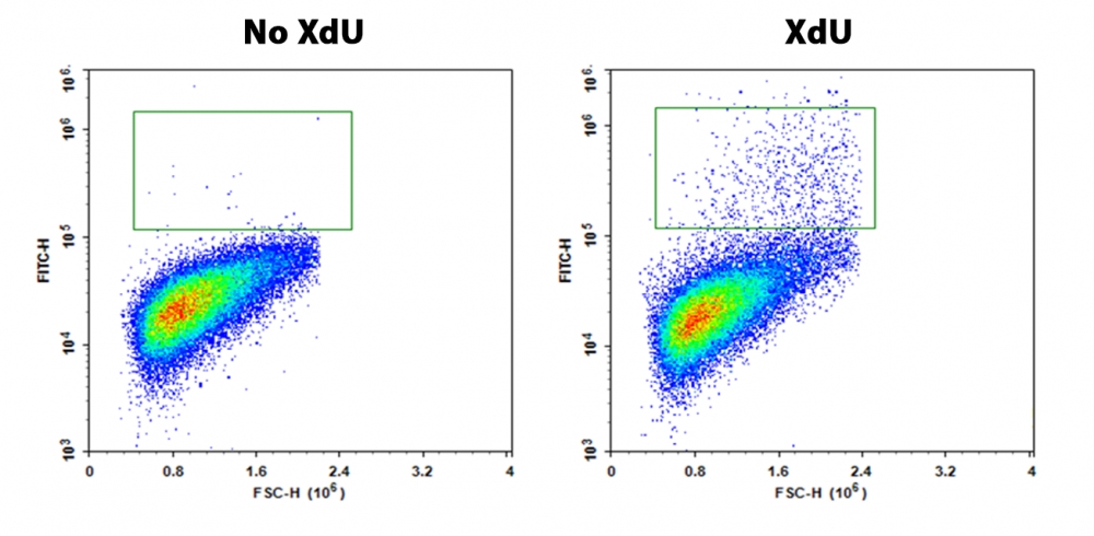
图1.用Buccutite XdU细胞增殖检测试剂盒(Cat#22323)检测到的S期Jurkat细胞的流式细胞仪中的反应。 将Jurkat细胞以50,000个细胞/孔/ 100μL的浓度接种在6孔黑色板上过夜。 按照说明书,将细胞在37℃下用XdU处理3小时,固定并透化。 然后将细胞在染色缓冲液中用iFluor 488叠氮化物染色30分钟,并用PBS洗涤3次。 使用FITC通道,通过NovoCyte流式细胞仪检测荧光响应。

说明书
Bucculite XdU细胞增殖检测试剂盒*蓝色激光兼容**流式细胞术*.pdf