Cell Meter 细胞活性检测试剂盒(活细胞/死细胞) *绿色/红色双重荧光* 货号22789-AAT Bioquest荧光染料

上海金畔生物科技有限公司代理AAT Bioquest荧光染料全线产品,欢迎访问AAT Bioquest荧光染料官网了解更多信息。

Cell Meter 细胞活性检测试剂盒(活细胞/死细胞) *绿色/红色双重荧光*

Cell Meter 细胞活性检测试剂盒(活细胞/死细胞) *绿色/红色双重荧光*

Cell Meter 细胞活性检测试剂盒(活细胞/死细胞) *绿色/红色双重荧光*    货号22789 货号 22789 存储条件 在零下15度以下保存, 避免光照
规格 200 Tests 价格 2604
Ex (nm) 492 Em (nm) 515
分子量 溶剂
产品详细介绍

简要概述

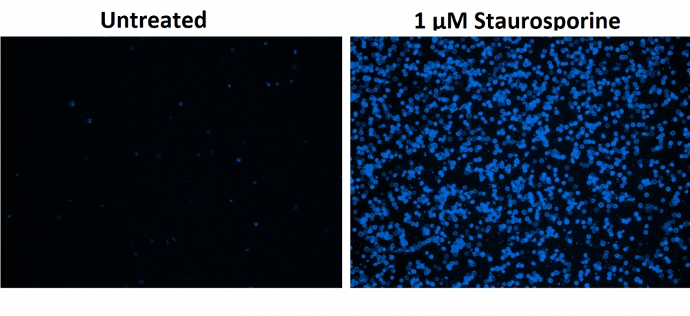
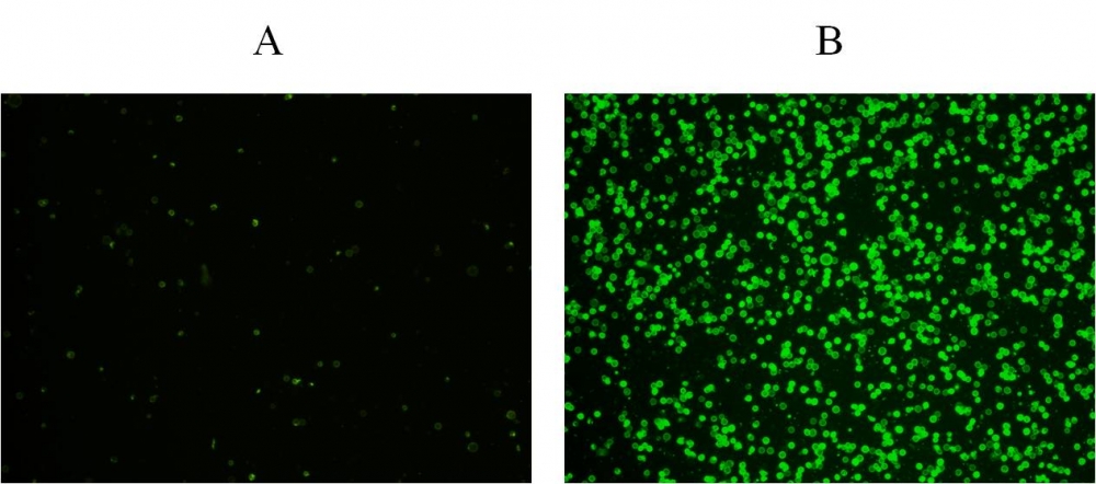
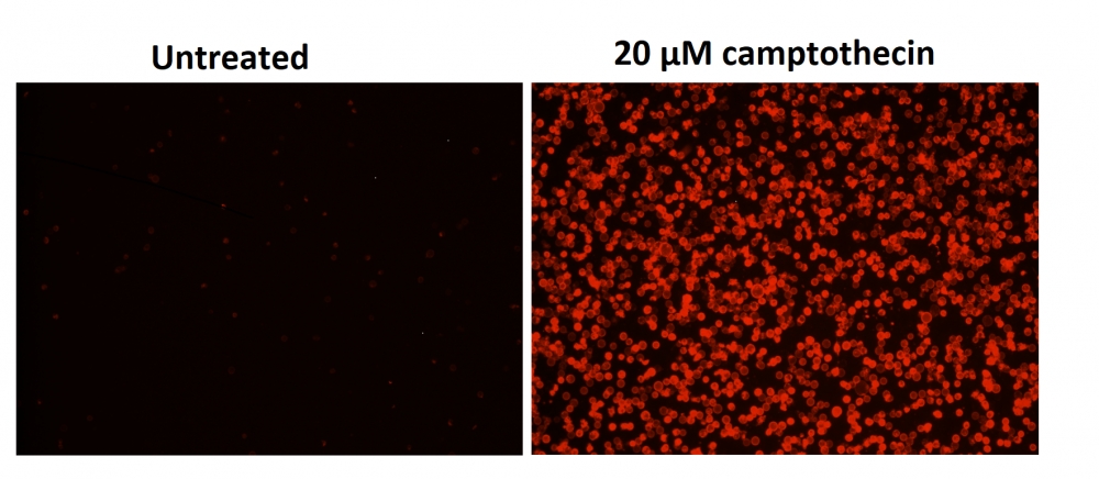
Cell Meter 细胞活性检测试剂盒是美国AAT Bioquest生产的细胞活性检测试剂盒,Cell Meter检测试剂盒是一类用于检测细胞功能的系列工具,包括细胞活性、细胞毒性、细胞凋亡、细胞膜电位以及细胞周期等方面的指标。每种检测方案均能提供不同荧光颜色的检测方案。这些高效的检测方案为从多角度研究细胞功能活动提供了一种十分有效的方法。Cell Meter细胞活力检测试剂盒 *绿色/红色双重荧光* 使用两种非荧光性指示剂:Calcein AM,用于活细胞;一种非细胞渗透性的DNA结合染料,用于细胞膜不完整的死细胞。Calcein AM是一种疏水性复合物,可以轻易地渗透进入完整的活细胞,通过酯酶的水解作用,产生强烈的荧光。通过细胞内酯酶水解非荧光性Calcein AM,形成具有强烈荧光的亲水性Calcein,并且保留在细胞质中。这种酯酶的活性与活细胞数量成比例关系。DNA结合染料极性非常大,不能渗透进入具有完整细胞膜的活细胞,它只有在与死细胞的DNA结合的情况下才发出荧光。生长在培养板中的细胞可以被染料,且在不到两小时内就可以完成定量分析。本检测法比其它活细胞检测法更加强大、稳定。它可以用于许多荧光实验平台的高通量分析中,例如微孔板分析,免疫组化和流式细胞术。本试剂盒提供所有必需组分和最佳检测方案。它适合于增殖型和非增殖型细胞,也可用于悬浮和贴壁细胞。96微孔板中,每孔用试剂100ul,本试剂盒提供的试剂足以进行100次检测;384微孔板中,每孔加试剂25ul,本试剂盒提供的试剂足以进行400次检测。金畔生物是AAT Bioquest的中国代理商,为您提供最优质的Cell Meter 细胞活性检测试剂盒。 


适用仪器


流式细胞仪  
激发: 488nm激光
发射: 530/30 nm, 610/20 nm滤波片
通道: FITC, PE-Texas Red通道
荧光显微镜  
激发: FITC(活),TRITC(死)
发射: FITC(活),TRITC(死)
推荐孔板: 黑色透明
荧光酶标仪  
激发: FITC(活),TRITC(死)
发射: FITC(活),TRITC(死)
cutoff: 515 nm, 590 nm
推荐孔板: 黑色孔板

产品说明书

样品实验方案

简要概述

1.用测试化合物制备细胞
2.加入相同体积的CytoCalcein Green / Propidium Iodide染料加工溶液(100μL/孔/ 96孔板或25μL/孔/ 384孔板)
3.在室温或37°C孵育1小时
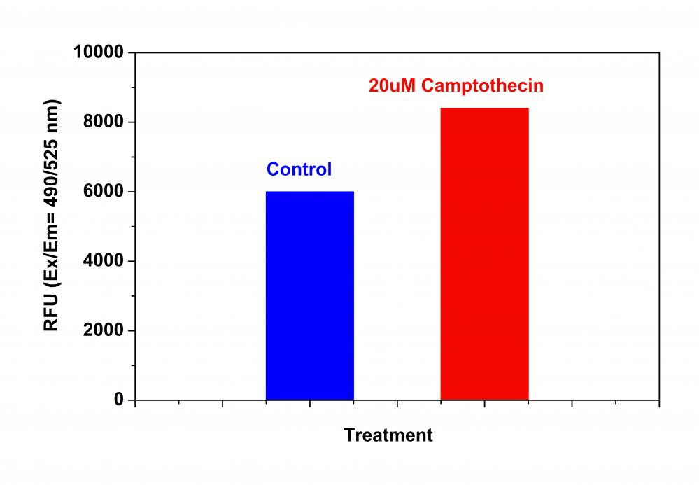
4.在强度下监测荧光(底部读取模式)Ex / Em = 490 / 525nm(截止= 515nm)和540 / 620nm(截止= 590nm),带有FITC滤光片(细胞存活)的荧光显微镜和TRITC滤光片 (细胞死亡),或FL1和FL2通道的流式细胞仪。


溶液配制

1.储存溶液配制

        除非另有说明,否则所有未使用的储备溶液应分成一次性等分试样,并在制备后储存在-20°C。 避免反复冻融循环。
CytoCalcein 绿色原液:
将20μLDMSO(组分C)加入到CytoCalcein Green(组分A)的小瓶中并充分混合以制备CytoCalcein Green原液。 避光。 注意:20μL的CytoCalcein Green原液足以用于一个平板。 存放时,密封管。


2.工作溶液配制

将全部内容物(20μL)的CytoCalcein Green储备溶液和20μL碘化丙啶(组分B)加入10mL测定缓冲液(组分C)中并充分混合以制备CytoCalcein Green /碘化丙锭染料 – 工作溶液。 CytoCalcein Green / Propidium Iodide染料加工溶液在室温下稳定至少2小时。 注意:如果CHO细胞等细胞含有导致荧光染料随时间泄漏的有机阴离子转运蛋白,应制备丙磺舒储备溶液,并加入到加样缓冲液中,最终孔内工作浓度范围为1到2.5 mM。 未使用的丙磺舒储备溶液可以在≤-20℃下储存。 由于最佳染色条件可能因细胞类型的不同而异,因此建议单独确定组分A和B的适当浓度。有关细胞样品制备的指南,请点击查看。


操作步骤

1.用酶标仪或荧光显微镜进行细胞活力测定:

1.1根据需要用测试化合物处理细胞。注意:在添加化合物之前不必洗涤细胞。然而,如果测试的化合物对血清敏感,则可在添加化合物之前吸出生长培养基和血清因子。在抽吸后加入100μL/孔/ 96孔板和25μL/孔/ 384孔板的1X Hank盐溶液和20mM Hepes缓冲液(HHBS)或您选择的缓冲液。或者,细胞可以在无血清培养基中生长。

1.2加入100μL/孔(96孔板)或25μL/孔(384孔板)的CytoCalcein TM Green / Propidium Iodide染料加工溶液。

1.3将板在室温或37°C孵育30分钟至1小时,避光。 (孵育时间可以从15分钟到过夜。我们得到了最佳结果,孵育时间少于4小时)。注意:适当的孵育时间取决于所使用的单个细胞类型和细胞浓度。优化每个实验的孵育时间。装载后请勿清洗细胞。对于非粘附细胞,建议在孵育后以800rpm离心细胞板2分钟,然后关闭制动器。

1.4使用荧光酶标仪(底部读取模式)在Ex / Em = 490 / 525nm(截止= 515nm)和Ex / Em = 540 / 620nm(截止= 590nm,细胞死亡)或荧光下监测荧光强度用于活细胞的FITC过滤器或用于死细胞的TRITC过滤器的显微镜。


2.使用流式细胞仪进行细胞活力测定:

2.1用测试化合物处理细胞一段所需的时间。

2.1离心细胞,得到1-5×10⁵个细胞/管。

2.3将细胞重悬于500μL的CytoCalcein™Green / Propidium Iodide染料加工溶液中。

2.4在室温或37°C孵育10至30分钟,避光。

可选:用HHBS或您选择的缓冲液清洗细胞。 将细胞重悬于500μLHHBS中,每管加入1-5×10⁵个细胞。

2.5用流式细胞仪在Ex / Em = 490 / 525nm和Ex / Em = 490 / 620nm(FL1和FL2通道)下监测荧光强度。


数据分析

Cell Meter 细胞活性检测试剂盒(活细胞/死细胞) *绿色/红色双重荧光*    货号22789

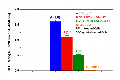
图1.使用Cell Meter Cell Viability Assay Kit测量Jurkat细胞对皂苷诱导的细胞死亡的影响。 用或不用0.5%皂苷处理2×10 6个细胞/ mL的Jurkat细胞5分钟。 离心细胞,用新鲜培养基替换上清液。 将100uL未处理的细胞(A),各50μL未处理和处理的细胞(B),25uL未处理的和75uL处理的细胞(C)和100uL的0.5%皂苷处理的细胞(D)接种在 96孔黑色墙壁/透明底部聚-D-赖氨酸板。 将细胞与100μL/孔的CytoCalcein Green / Propidium Iodide染料 – 工作溶液在37℃下孵育1小时。 使用NOVOstar仪器(BMG Labtech)在底部读取模式下在Ex / Em = 490 / 525nm和540 / 650nm下测量荧光强度。 如所示(n = 6)显示活细胞和死细胞上490 / 525nm与540 / 650nm荧光强度的比率。


参考文献

Effect of copper nanoparticles on physico-chemical properties of chitosan and gelatin-based scaffold developed for skin tissue engineering application
Authors: Shikha Kumari, Bhisham Narayan Singh, Pradeep Srivastava
Journal: 3 Biotech (2019): 102

Functional imaging of neuronal activity of auditory cortex by using Cal-520 in anesthetized and awake mice
Authors: Jingcheng Li, Jianxiong Zhang, Meng Wang, Junxia Pan, Xiaowei Chen, Xiang Liao
Journal: Biomedical Optics Express (2017): 2599–2610

NINJ2–A novel regulator of endothelial inflammation and activation
Authors: Jingjing Wang, Jingjing Fa, Pengyun Wang, Xinzhen Jia, Huixin Peng, Jing Chen, Yifan Wang, Chenhui Wang, Qiuyun Chen, Xin Tu
Journal: Cellular Signalling (2017)

Erythropoietin Stimulates Endothelial Progenitor Cells to Induce Endothelialization in an Aneurysm Neck After Coil Embolization by Modulating Vascular Endothelial Growth Factor
Authors: Peixi Liu, Yingjie Zhou, Qingzhu An, Yaying Song, Xi Chen, Guo-Yuan Yang, Wei Zhu
Journal: MEDICINE (2016): 1–8

Flexible Endoscopic Spray Application of Respiratory Epithelial Cells as Platform Technology to Apply Cells in Tubular Organs
Authors: Anja Lena Thiebes, Manuel Armin Reddemann, Johannes Palmer, Reinhold Kneer, Stefan Jockenhoevel, Christian Gabriel Cornelissen
Journal: Tissue Engineering Part C: Methods (2016): 322–331

Influence of hypothermia and subsequent rewarming upon leukocyte-endothelial interactions and expression of Junctional-Adhesion-Molecules A and B
Authors: Nicolai V Bogert, Isabella Werner, Angela Kornberger, Patrick Meybohm, Anton Moritz, Till Keller, Ulrich A Stock, Andres Beiras-Fernandez
Journal: Scientific reports (2016)

Inhibition of ABC transport proteins by oil sands process affected water
Authors: Hattan A Alharbi, David MV Saunders, Ahmed Al-Mousa, Jane Alcorn, Alberto S Pereira, Jonathan W Martin, John P Giesy, Steve B Wiseman
Journal: Aquatic Toxicology (2016): 81–88

Rapid generation of collagen-based microtissues to study cell–matrix interactions
Authors: Marie-Elena Brett, Alexandra L Crampton, David K Wood
Journal: Technology (2016): 1–8

Toxicokinetics and toxicodynamics of chlorpyrifos is altered in embryos of Japanese medaka exposed to oil sands process-affected water: evidence for inhibition of P-glycoprotein
Authors: Hattan A Alharbi, Jane Alcorn, Ahmed Al-Mousa, John P Giesy, Steve B Wiseman
Journal: Journal of Applied Toxicology (2016)

Spraying respiratory epithelial cells to coat tissue-engineered constructs
Authors: Anja Lena Thiebes, Stefanie Albers, Christian Klopsch, Stefan Jockenhoevel, Christian G Cornelissen
Journal: BioResearch open access (2015): 278–287


相关产品

产品名称 货号
Live or Dead 细胞活性检测试剂盒 *红/蓝双色荧光* Cat#22788

说明书
Cell Meter 细胞活性检测试剂盒(活细胞/死细胞) *绿色/红色双重荧光*.pdf

层析柱,具内螺纹口,欣维尔,C432801

上海金畔生物科技有限公司提供层析柱,具内螺纹口,欣维尔,C432801,可以访问官网了解更多产品信息。
层析柱,具内螺纹口,欣维尔,C432801

询价
欣维尔

技术参数

层析柱,具内螺纹口,欣维尔,C432801

*注意:尽管我们采用的是厚壁玻璃材质,其安全系数比一般层析柱大大
加强,我们仍然推荐您在通风橱内操作此产品,并关上通风橱的玻璃门。

材质 玻璃
规格(mm) 203
标准口 #25
砂芯
活塞

多肽定制Parallel topology beta-Amyloid modified peptide 编码

上海金畔生物科技有限公司可以定制不同序列多肽,可以访问官网了解更多产品信息。

名称 Parallel topology beta-Amyloid modified peptide
编码
别名 Parallel topology beta-Amyloid modified peptide
纯度 80%,90%,95%,98%,99%
重量 1mg,5mg,10mg,50mg,100mg,1g
序列(单字母缩写) CHQKLVFFAEDYNGKDEAFFVLKQHW
序列(三字母缩写) Cys-His-Gln-Lys-Leu-Val-Phe-Phe-Ala-Glu-Asp-Tyr-Asn-Gly-Lys-Asp-Glu-Ala-Phe-Phe-Val-Leu-Lys-Gln-His-Trp
基本描述
溶解度
分子量 3199.65
化学式 C151H211N37O39S
存储条件 Store at -20°C. Keep tightly closed. Store in a cool dry place.
注释
Documents Parallel topology beta-Amyloid modified peptide          编码
Figures Parallel topology beta-Amyloid modified peptide          编码
Reference
C端
N端
化学桥

Cell Meter 磷脂酰丝氨酸凋亡检测试剂盒 蓝色荧光,适合微孔板检测 货号22790-AAT Bioquest荧光染料

上海金畔生物科技有限公司代理AAT Bioquest荧光染料全线产品,欢迎访问AAT Bioquest荧光染料官网了解更多信息。

Cell Meter 磷脂酰丝氨酸凋亡检测试剂盒 蓝色荧光,适合微孔板检测

Cell Meter 磷脂酰丝氨酸凋亡检测试剂盒 蓝色荧光,适合微孔板检测

Cell Meter 磷脂酰丝氨酸凋亡检测试剂盒 蓝色荧光,适合微孔板检测     货号22790 货号 22790 存储条件 在2-8度冷藏保存, 避免光照
规格 100 Tests 价格 2604
Ex (nm) 405 Em (nm) 450
分子量 溶剂
产品详细介绍

简要概述

我们的Cell Meter™检测试剂盒是一套用于监测细胞活性的试剂盒。有多种参数可用于监测细胞活力。该特定试剂盒旨在通过测量磷脂酰丝氨酸(PS)的转运来监测细胞凋亡。在细胞凋亡中,PS转移到质膜的外部小叶。磷脂酰丝氨酸在细胞表面的出现是细胞凋亡的初始/中间阶段的通用指标,可以在观察形态变化之前进行检测。该特定试剂盒旨在通过测量磷脂酰丝氨酸(PS)的转运来监测细胞凋亡。该试剂盒使用我们专有的基于荧光小分子的Apopxin™PS探针,该探针以比膜联蛋白V(Kd <10 nM)高得多的亲和力特异性结合PS。与膜PS结合后具有蓝色荧光。它可以以酶标仪,显微镜和流式细胞仪的形式使用,而大多数其他商业凋亡检测试剂盒只能与显微镜或流式细胞仪平台一起使用。


适用仪器


荧光酶标仪  
激发: 405nm
发射: 450nm
cutoff: 420nm
推荐孔板: 黑色孔板
读取模式: 底读模式

产品说明书

样品实验方案

简要概述

1.用测试化合物制备细胞(100 µL /孔/ 96孔板或25 µL /孔/ 384孔板)
2.加入等量的Apopxin™Violet 450工作溶液
3.在室温下孵育1小时
4.观察Ex / Em = 405/450 nm(截止= 420 nm)的荧光强度(底部读取模式)或使用装有紫罗兰色滤光片的荧光显微镜


溶液配制

工作溶液配制

将10μL100X Apopxin™紫罗兰色450(组分A)添加到1 mL的测定缓冲液(组分B)中,并充分混合以制成Apopxin™紫罗兰色450工作溶液。 


操作步骤

1.通过向PBS或所需的缓冲液中加入10X /孔(96孔板)或2.5 µL /孔(384孔板)的10X测试化合物储备溶液来处理细胞。对于空白孔(不含细胞的培养基),添加相同量的化合物缓冲液。

2.将细胞板在5%CO2、37°C​​的培养箱中孵育所需的时间(对于用星形孢菌素处理的Jurkat细胞需要4-6小时)以诱导凋亡。注意:由于在405 nm激发时,从380至490 nm的宽发射光谱,某些化合物(例如喜树碱)可能会产生假阳性反应。

3.在每个孔中添加100 µL /孔(96孔板)或25 µL /孔(384孔板)的Apopxin™Violet 450工作溶液。

4.在避光条件下,于室温下孵育细胞板至少1小时。

5.以800 rpm的速度离心细胞板(特别是非粘附细胞)2分钟(制动)。

6.使用荧光酶标仪(底部读取模式)在Ex / Em = 405/450 nm(截止= 420 nm)或使用带有紫色滤光片的荧光显微镜对图像池进行荧光强度监测。

Cell Meter 磷脂酰丝氨酸凋亡检测试剂盒 蓝色荧光,适合微孔板检测     货号22790

图1.用Apopxin™Violet 450偶联物染色的HeLa细胞的荧光图像。 Jurkat细胞在37℃下不使用(左)或用1μM星形孢菌素(右)处理4小时。 使用带有紫色滤光片组的显微镜(激发= 405nm)测量荧光强度。

参考文献

Physiological effects of the herbicide glyphosate on the cyanobacterium Microcystis aeruginosa
Authors: Wu, Liang and Qiu, Zhihao and Zhou, Ya and Du, Yuping and Liu, Chaonan and Ye, Jing and Hu, Xiaojun
Journal: Aquatic Toxicology (2016): 72–79

Tumor-selective mitochondrial network collapse induced by atmospheric gas plasma-activated medium.
Authors: Saito, Kosuke and Asai, Tomohiko and Fujiwara, Kyoko and Sahara, Junki and Koguchi, Haruhisa and Fukuda, Noboru and Suzuki-Karasaki, Miki and Soma, Masayoshi and Suzuki-Karasaki, Yoshihiro
Journal: Oncotarget (2016)

Inhibition of malignant phenotypes of human osteosarcoma cells by a gene silencer, a pyrrole–imidazole polyamide, which targets an E-box motif
Authors: Taniguchi, Masashi and Fujiwara, Kyoko and Nakai, Yuji and Ozaki, Toshinori and Koshikawa, Nobuko and Toshio, Kojima and Kataba, Motoaki and Oguni, Asako and Matsuda, Hiroyuki and Yoshida, Yukihiro and others
Journal: FEBS open bio (2014): 328–334

Detection of apoptosis based on the interaction between annexin V and phosphatidylserine
Authors: Liu T, Zhu W, Yang X, Chen L, Yang R, Hua Z, Li G.
Journal: Anal Chem (2009): 2410

Evaluation of cell surface expression of phosphatidylserine in ovarian carcinoma effusions using the annexin-V/7-AAD assay: clinical relevance and comparison with other apoptosis parameters
Authors: Dong HP, Holth A, Kleinberg L, Ruud MG, Elstr and MB, Trope CG, Davidson B, Risberg B.
Journal: Am J Clin Pathol (2009): 756

Mobilization of lysosomal calcium regulates the externalization of phosphatidylserine during apoptosis
Authors: Mirnikjoo B, Balasubramanian K, Schroit AJ.
Journal: J Biol Chem (2009): 6918

Peptidic targeting of phosphatidylserine for the MRI detection of apoptosis in atherosclerotic plaques
Authors: Burtea C, Laurent S, Lancelot E, Ballet S, Murariu O, Rousseaux O, Port M, V and er Elst L, Corot C, Muller RN.
Journal: Mol Pharm (2009): 1903

Suicidal membrane repair regulates phosphatidylserine externalization during apoptosis
Authors: Mirnikjoo B, Balasubramanian K, Schroit AJ.
Journal: J Biol Chem (2009): 22512

Trivalent methylated arsenical-induced phosphatidylserine exposure and apoptosis in platelets may lead to increased thrombus formation
Authors: Bae ON, Lim KM, Noh JY, Chung SM, Kim SH, Chung JH.
Journal: Toxicol Appl Pharmacol (2009): 144

Discovery of a phosphatidylserine-recognizing peptide and its utility in molecular imaging of tumour apoptosis
Authors: Thapa N, Kim S, So IS, Lee BH, Kwon IC, Choi K, Kim IS.
Journal: J Cell Mol Med (2008): 1649

说明书
Cell Meter 磷脂酰丝氨酸凋亡检测试剂盒 蓝色荧光,适合微孔板检测 .pdf

多肽定制Parasin I 编码 [219552-69-9]

上海金畔生物科技有限公司可以定制不同序列多肽,可以访问官网了解更多产品信息。

名称 Parasin I
编码 [219552-69-9]
别名 Parasin I
纯度 80%,90%,95%,98%,99%
重量 1mg,5mg,10mg,50mg,100mg,1g
序列(单字母缩写) KGRGKQGGKVRAKAKTRSS
序列(三字母缩写) Lys-Gly-Arg-Gly-Lys-Gln-Gly-Gly-Lys-Val-Arg-Ala-Lys-Ala-Lys-Thr-Arg-Ser-Ser
基本描述
溶解度
分子量 2000.36
化学式 C82H154N34O24
存储条件 Store at -20°C. Keep tightly closed. Store in a cool dry place.
注释
Documents Parasin I           编码     [219552-69-9]
Figures Parasin I           编码     [219552-69-9]
Reference
C端
N端
化学桥

层析柱,具内螺纹口,欣维尔,C432605

上海金畔生物科技有限公司提供层析柱,具内螺纹口,欣维尔,C432605,可以访问官网了解更多产品信息。
层析柱,具内螺纹口,欣维尔,C432605

询价
欣维尔

技术参数

层析柱,具内螺纹口,欣维尔,C432605

*注意:尽管我们采用的是厚壁玻璃材质,其安全系数比一般层析柱大大
加强,我们仍然推荐您在通风橱内操作此产品,并关上通风橱的玻璃门。

材质 玻璃
规格(mm) 610
标准口 #25
砂芯
活塞

Cell Meter 磷脂酰丝氨酸凋亡检测试剂盒 绿色荧光,适合微孔板检测 货号22791-AAT Bioquest荧光染料

上海金畔生物科技有限公司代理AAT Bioquest荧光染料全线产品,欢迎访问AAT Bioquest荧光染料官网了解更多信息。

Cell Meter 磷脂酰丝氨酸凋亡检测试剂盒 绿色荧光,适合微孔板检测

Cell Meter 磷脂酰丝氨酸凋亡检测试剂盒 绿色荧光,适合微孔板检测

Cell Meter 磷脂酰丝氨酸凋亡检测试剂盒 绿色荧光,适合微孔板检测     货号22791 货号 22791 存储条件 在2-8度冷藏保存, 避免光照
规格 100 Tests 价格 2604
Ex (nm) 498 Em (nm) 520
分子量 溶剂
产品详细介绍

简要概述

Cell Meter检测试剂盒是一类用于检测细胞功能的系列工具,包括细胞活性、细胞毒性、细胞凋亡、细胞膜电位以及细胞周期等方面的指标。每种检测方案均能提供不同荧光颜色的检测方案。这些高效的检测方案为从多角度研究细胞功能活动提供了一种十分有效的方法。Cell Meter磷脂酰丝氨酸凋亡检测试剂盒 *绿色荧光,适合微孔板检测*是通过检测PS的翻转来测定细胞凋亡。在凋亡细胞中,PS从胞内移位到胞外表面,因此PS出现在细胞的外表面可以作为细胞凋亡的起始或者中间阶段的通用指示器,也可以通过其形态学改变来测定。该试剂盒使用我们独有的绿色荧光Apopxin PS传感器特异性的结合PS,其亲和性高于Annexin V(Kd<10nM)。该试剂盒使用的PS传感器结合膜上的PS后,发出绿色的荧光。因为与PS的高亲和性,该试剂盒比其它基于Annexin-V,仅能用荧光显微镜和流式检测凋亡的试剂盒更稳定,并且本试剂盒除上述两种仪器外还能使用荧光酶标仪。

点击查看细胞样品制备


适用仪器


荧光酶标仪  
激发: 490nm
发射: 525nm
cutoff: 515nm
推荐孔板: 黑色透明
读取模式: 底读模式

产品说明书

样品实验方案

简要概述

1.用测试化合物制备细胞(100 µL /孔/ 96孔板或25 µL /孔/ 384孔板)
2.加入等量的Apopxin™Green工作溶液
3.在室温下孵育1小时
4.在Ex / Em = 490/525 nm(截止= 515 nm)或带有FITC滤光片的荧光显微镜下监控荧光强度(底部读取模式)


溶液配制

工作溶液配制

将10 µL 100X Apopxin™Green(组分A)添加到1 mL的测定缓冲液(组分B)中,并充分混合以制成Apopxin™Green工作溶液。


操作步骤

1.通过向细胞制备缓冲液中加入10XL /孔(96孔板)的10X测试化合物溶液,以所需的测试化合物处理细胞,总体积为100μL/孔。对于384孔板,请在细胞制备缓冲液中使用5 µL /孔的5X测试化合物溶液,总体积为25 µL /孔。对于空白孔(不含细胞的培养基),添加相同量的化合物缓冲液。

2.将细胞板在5%CO2、37°C​​的培养箱中孵育所需的时间(对于喜树碱处理过的Jurkat细胞,需要4-6小时)以诱导凋亡。

3.在每个孔中添加100 µL /孔(96孔板)或25 µL /孔(384孔板)的Apopxin™Green工作溶液。

4.在避光条件下,于室温下孵育细胞板至少1小时。

5.以800 rpm的速度离心细胞板(特别是非粘附细胞)2分钟(制动)。

6.使用荧光酶标仪(Ext / Em = 490/525 nm(Cutoff = 515 nm))或使用带有FITC滤光片的荧光显微镜的图像池监控荧光强度。


图示

Cell Meter 磷脂酰丝氨酸凋亡检测试剂盒 绿色荧光,适合微孔板检测     货号22791

图1.在Costar黑色壁/透明底部96孔板中,用Cell Meter™磷脂酰丝氨酸凋亡检测试剂盒染色的Jurkat细胞图像。 A:未经处理的对照细胞。 B:用20 µM喜树碱处理5小时的细胞。

参考文献

Physiological effects of the herbicide glyphosate on the cyanobacterium Microcystis aeruginosa
Authors: Wu, Liang and Qiu, Zhihao and Zhou, Ya and Du, Yuping and Liu, Chaonan and Ye, Jing and Hu, Xiaojun
Journal: Aquatic Toxicology (2016): 72–79

Tumor-selective mitochondrial network collapse induced by atmospheric gas plasma-activated medium.
Authors: Saito, Kosuke and Asai, Tomohiko and Fujiwara, Kyoko and Sahara, Junki and Koguchi, Haruhisa and Fukuda, Noboru and Suzuki-Karasaki, Miki and Soma, Masayoshi and Suzuki-Karasaki, Yoshihiro
Journal: Oncotarget (2016)

Inhibition of malignant phenotypes of human osteosarcoma cells by a gene silencer, a pyrrole–imidazole polyamide, which targets an E-box motif
Authors: Taniguchi, Masashi and Fujiwara, Kyoko and Nakai, Yuji and Ozaki, Toshinori and Koshikawa, Nobuko and Toshio, Kojima and Kataba, Motoaki and Oguni, Asako and Matsuda, Hiroyuki and Yoshida, Yukihiro and others
Journal: FEBS open bio (2014): 328–334

Detection of apoptosis based on the interaction between annexin V and phosphatidylserine
Authors: Liu T, Zhu W, Yang X, Chen L, Yang R, Hua Z, Li G.
Journal: Anal Chem (2009): 2410

Evaluation of cell surface expression of phosphatidylserine in ovarian carcinoma effusions using the annexin-V/7-AAD assay: clinical relevance and comparison with other apoptosis parameters
Authors: Dong HP, Holth A, Kleinberg L, Ruud MG, Elstr and MB, Trope CG, Davidson B, Risberg B.
Journal: Am J Clin Pathol (2009): 756

Mobilization of lysosomal calcium regulates the externalization of phosphatidylserine during apoptosis
Authors: Mirnikjoo B, Balasubramanian K, Schroit AJ.
Journal: J Biol Chem (2009): 6918

Peptidic targeting of phosphatidylserine for the MRI detection of apoptosis in atherosclerotic plaques
Authors: Burtea C, Laurent S, Lancelot E, Ballet S, Murariu O, Rousseaux O, Port M, V and er Elst L, Corot C, Muller RN.
Journal: Mol Pharm (2009): 1903

Suicidal membrane repair regulates phosphatidylserine externalization during apoptosis
Authors: Mirnikjoo B, Balasubramanian K, Schroit AJ.
Journal: J Biol Chem (2009): 22512

Trivalent methylated arsenical-induced phosphatidylserine exposure and apoptosis in platelets may lead to increased thrombus formation
Authors: Bae ON, Lim KM, Noh JY, Chung SM, Kim SH, Chung JH.
Journal: Toxicol Appl Pharmacol (2009): 144

Discovery of a phosphatidylserine-recognizing peptide and its utility in molecular imaging of tumour apoptosis
Authors: Thapa N, Kim S, So IS, Lee BH, Kwon IC, Choi K, Kim IS.
Journal: J Cell Mol Med (2008): 1649

说明书
Cell Meter 磷脂酰丝氨酸凋亡检测试剂盒 绿色荧光,适合微孔板检测 .pdf

多肽定制Parasin I 编码

上海金畔生物科技有限公司可以定制不同序列多肽,可以访问官网了解更多产品信息。

名称 Parasin I
编码
别名 Parasin I
纯度 80%,90%,95%,98%,99%
重量 1mg,5mg,10mg,50mg,100mg,1g
序列(单字母缩写) KGRGKQGGKVRAKAKTRSS-OH
序列(三字母缩写) Lys-Gly-Arg-Gly-Lys-Gln-Gly-Gly-Lys-Val-Arg-Ala-Lys-Ala-Lys-Thr-Arg-Ser-Ser
基本描述
溶解度
分子量 0
化学式
存储条件 Store at -20°C. Keep tightly closed. Store in a cool dry place.
注释
Documents Parasin I          编码
Figures Parasin I          编码
Reference
C端
N端
化学桥

层析柱,具内螺纹口,欣维尔,C432604

上海金畔生物科技有限公司提供层析柱,具内螺纹口,欣维尔,C432604,可以访问官网了解更多产品信息。
层析柱,具内螺纹口,欣维尔,C432604

询价
欣维尔

技术参数

层析柱,具内螺纹口,欣维尔,C432604

*注意:尽管我们采用的是厚壁玻璃材质,其安全系数比一般层析柱大大
加强,我们仍然推荐您在通风橱内操作此产品,并关上通风橱的玻璃门。

材质 玻璃
规格(mm) 457
标准口 #25
砂芯
活塞

Cell Meter 磷脂酰丝氨酸凋亡检测试剂盒 红色荧光,适合微孔板检测 货号22792-AAT Bioquest荧光染料

上海金畔生物科技有限公司代理AAT Bioquest荧光染料全线产品,欢迎访问AAT Bioquest荧光染料官网了解更多信息。

Cell Meter 磷脂酰丝氨酸凋亡检测试剂盒 红色荧光,适合微孔板检测

Cell Meter 磷脂酰丝氨酸凋亡检测试剂盒 红色荧光,适合微孔板检测

Cell Meter 磷脂酰丝氨酸凋亡检测试剂盒 红色荧光,适合微孔板检测     货号22792 货号 22792 存储条件 在2-8度冷藏保存, 避免光照
规格 100 Tests 价格 2604
Ex (nm) 575 Em (nm) 600
分子量 溶剂
产品详细介绍

简要概述

Cell Meter检测试剂盒是一类用于检测细胞功能的系列工具,包括细胞活性、细胞毒性、细胞凋亡、细胞膜电位以及细胞周期等方面的指标。每种检测方案均能提供不同荧光颜色的检测方案。这些高效的检测方案为从多角度研究细胞功能活动提供了一种十分有效的方法。Cell Meter磷脂酰丝氨酸凋亡检测试剂盒 *红色荧光,适合微孔板检测*是通过检测PS的翻转来测定细胞凋亡。在凋亡细胞中,PS从胞内移位到胞外表面,因此PS出现在细胞的外表面可以作为细胞凋亡的起始或者中间阶段的通用指示器,也可以通过其形态学改变来测定。该试剂盒使用我们独有的红色荧光Apopxin PS传感器特异性的结合PS,其亲和性高于Annexin V(Kd<10nM)。该试剂盒使用的PS传感器结合膜上的PS后,发出红色的荧光。因为与PS的高亲和性,该试剂盒比其它基于Annexin-V,仅能用荧光显微镜和流式检测凋亡的试剂盒更稳定,并且本试剂盒除上述两种仪器外还能使用荧光酶标仪。

点击查看细胞样品配制


适用仪器


荧光酶标仪  
激发: 590nm
发射: 630nm
cutoff: 610nm
推荐孔板: 黑色透明
读取模式: 底读模式

产品说明书

样品实验方案

简要概述

1.用测试化合物制备细胞(100 µL /孔/ 96孔板或25 µL /孔/ 384孔板)
2.加入等量的Apopxin™Red工作溶液
3.在室温下孵育1小时
4.监视Ex / Em = 590/630 nm(截止= 610 nm)的荧光强度(底部读取模式)或使用带有Texas Red滤光片的荧光显微镜


溶液配制

工作溶液配制

将10 µL Apopxin™Red(组分A)加入1 mL的测定缓冲液(组分B)中,并充分混合以制成Apopxin™Red工作溶液。


操作步骤

1.通过向PBS或所需的缓冲液中加入10X /孔(96孔板)或2.5 µL /孔(384孔板)的10X测试化合物储备溶液来处理细胞。 对于空白孔(不含细胞的培养基),添加相同量的化合物缓冲液。

2.将细胞板在5%CO2、37°C的培养箱中孵育所需的时间(对于喜树碱处理过的Jurkat细胞,需要4-6小时)以诱导凋亡。

3.在每个孔中加入100 µL /孔(96孔板)或25 µL /孔(384孔板)的Apopxin™Red工作溶液。

4.在避光条件下,于室温下孵育细胞板至少1小时。

5.以800 rpm的速度离心细胞板(特别是非粘附细胞)2分钟(制动)。

6.使用荧光酶标仪(Ext / Em = 590/630 nm(截止= 610 nm))或使用带有TexasRed®滤光片的荧光显微镜对图像池进行荧光强度监测。


图示

Cell Meter 磷脂酰丝氨酸凋亡检测试剂盒 红色荧光,适合微孔板检测     货号22792

图1.用Cell Meter™磷脂酰丝氨酸凋亡检测试剂盒染色的Costar黑壁/透明底部96孔板中Jurkat细胞的荧光图像。 不使用(左)或用20μM喜树碱(右)处理Jurkat细胞5小时。 使用具有Texas Red通道的荧光显微镜测量荧光强度。


参考文献

Physiological effects of the herbicide glyphosate on the cyanobacterium Microcystis aeruginosa
Authors: Wu, Liang and Qiu, Zhihao and Zhou, Ya and Du, Yuping and Liu, Chaonan and Ye, Jing and Hu, Xiaojun
Journal: Aquatic Toxicology (2016): 72–79

Tumor-selective mitochondrial network collapse induced by atmospheric gas plasma-activated medium.
Authors: Saito, Kosuke and Asai, Tomohiko and Fujiwara, Kyoko and Sahara, Junki and Koguchi, Haruhisa and Fukuda, Noboru and Suzuki-Karasaki, Miki and Soma, Masayoshi and Suzuki-Karasaki, Yoshihiro
Journal: Oncotarget (2016)

Inhibition of malignant phenotypes of human osteosarcoma cells by a gene silencer, a pyrrole–imidazole polyamide, which targets an E-box motif
Authors: Taniguchi, Masashi and Fujiwara, Kyoko and Nakai, Yuji and Ozaki, Toshinori and Koshikawa, Nobuko and Toshio, Kojima and Kataba, Motoaki and Oguni, Asako and Matsuda, Hiroyuki and Yoshida, Yukihiro and others
Journal: FEBS open bio (2014): 328–334

Detection of apoptosis based on the interaction between annexin V and phosphatidylserine
Authors: Liu T, Zhu W, Yang X, Chen L, Yang R, Hua Z, Li G.
Journal: Anal Chem (2009): 2410

Evaluation of cell surface expression of phosphatidylserine in ovarian carcinoma effusions using the annexin-V/7-AAD assay: clinical relevance and comparison with other apoptosis parameters
Authors: Dong HP, Holth A, Kleinberg L, Ruud MG, Elstr and MB, Trope CG, Davidson B, Risberg B.
Journal: Am J Clin Pathol (2009): 756

Mobilization of lysosomal calcium regulates the externalization of phosphatidylserine during apoptosis
Authors: Mirnikjoo B, Balasubramanian K, Schroit AJ.
Journal: J Biol Chem (2009): 6918

Peptidic targeting of phosphatidylserine for the MRI detection of apoptosis in atherosclerotic plaques
Authors: Burtea C, Laurent S, Lancelot E, Ballet S, Murariu O, Rousseaux O, Port M, V and er Elst L, Corot C, Muller RN.
Journal: Mol Pharm (2009): 1903

Suicidal membrane repair regulates phosphatidylserine externalization during apoptosis
Authors: Mirnikjoo B, Balasubramanian K, Schroit AJ.
Journal: J Biol Chem (2009): 22512

Trivalent methylated arsenical-induced phosphatidylserine exposure and apoptosis in platelets may lead to increased thrombus formation
Authors: Bae ON, Lim KM, Noh JY, Chung SM, Kim SH, Chung JH.
Journal: Toxicol Appl Pharmacol (2009): 144

Discovery of a phosphatidylserine-recognizing peptide and its utility in molecular imaging of tumour apoptosis
Authors: Thapa N, Kim S, So IS, Lee BH, Kwon IC, Choi K, Kim IS.
Journal: J Cell Mol Med (2008): 1649

说明书
Cell Meter 磷脂酰丝氨酸凋亡检测试剂盒 红色荧光,适合微孔板检测 .pdf

多肽定制Parathyroid Hormone (1-13) 编码

上海金畔生物科技有限公司可以定制不同序列多肽,可以访问官网了解更多产品信息。

名称 Parathyroid Hormone (1-13)
编码
别名 Parathyroid Hormone (1-13)
纯度 80%,90%,95%,98%,99%
重量 1mg,5mg,10mg,50mg,100mg,1g
序列(单字母缩写) SVSEIQLMHNLGK
序列(三字母缩写) H-Ser-Val-Ser-Glu-Ile-Gln-Leu-Met-His-Asn-Leu-Gly-Lys-OH (trifluoroacetate salt)
基本描述 Parathyroid hormone (PTH) regulates the metabolism of calcium and phosphate. PTH and PTH-related polypeptide (PTHrP) play important roles in calcium homeostasis of bone, kidney, breast, and placenta; they signal via the PTH/PTHrP and PTH2 receptors. PTH(1?34) administration suppresses cardiovascular calcification and down-regulates aortic osteogenic programs driven by diabetes and dyslipidemia.
溶解度
分子量 1455.7
化学式 C62H106N18O20S1
存储条件 Store at -20°C. Keep tightly closed. Store in a cool dry place.
注释
Documents Parathyroid Hormone (1-13)          编码
Figures Parathyroid Hormone (1-13)          编码
Reference Kumar, V. et al. Clin Chem 56, 306 (2010) Lopez, M. et al. Clin Chem 56, 281 (2010).
C端
N端
化学桥

层析柱,具内螺纹口,欣维尔,C432603

上海金畔生物科技有限公司提供层析柱,具内螺纹口,欣维尔,C432603,可以访问官网了解更多产品信息。
层析柱,具内螺纹口,欣维尔,C432603

询价
欣维尔

技术参数

层析柱,具内螺纹口,欣维尔,C432603

*注意:尽管我们采用的是厚壁玻璃材质,其安全系数比一般层析柱大大
加强,我们仍然推荐您在通风橱内操作此产品,并关上通风橱的玻璃门。

材质 玻璃
规格(mm) 305
标准口 #25
砂芯
活塞

多肽定制Parathyroid Hormone (13-34), human 编码 [81306-64-1]

上海金畔生物科技有限公司可以定制不同序列多肽,可以访问官网了解更多产品信息。

名称 Parathyroid Hormone (13-34), human
编码 [81306-64-1]
别名 Parathyroid Hormone (13-34), human
纯度 80%,90%,95%,98%,99%
重量 1mg,5mg,10mg,50mg,100mg,1g
序列(单字母缩写) KHLNSMERVEWLRKKLQDVHNF
序列(三字母缩写) Lys-His-Leu-Asn-Ser-Met-Glu-Arg-Val-Glu-Trp-Leu-Arg-Lys-Lys-Leu-Gln-Asp-Val-His-Asn-Phe
基本描述
溶解度
分子量 2808.26
化学式 C125H199N39O33S
存储条件 Store at -20°C. Keep tightly closed. Store in a cool dry place.
注释
Documents Parathyroid Hormone (13-34), human           编码     [81306-64-1]
Figures Parathyroid Hormone (13-34), human           编码     [81306-64-1]
Reference
C端
N端
化学桥

Cell Meter Caspase 8活性细胞凋亡检测试剂盒 绿色荧光 货号22798-AAT Bioquest荧光染料

上海金畔生物科技有限公司代理AAT Bioquest荧光染料全线产品,欢迎访问AAT Bioquest荧光染料官网了解更多信息。

Cell Meter Caspase 8活性细胞凋亡检测试剂盒 绿色荧光

Cell Meter Caspase 8活性细胞凋亡检测试剂盒 绿色荧光

Cell Meter Caspase 8活性细胞凋亡检测试剂盒 绿色荧光     货号22798 货号 22798 存储条件 在零下15度以下保存, 避免光照
规格 200 Tests 价格 2604
Ex (nm) 500 Em (nm) 522
分子量 溶剂
产品详细介绍

简要概述

Cell Meter Caspase 8活性细胞凋亡检测试剂盒是美国AAT Bioquest生产的用于细胞凋亡的试剂盒,Cell Meter Caspase 8活性细胞凋亡检测试剂盒是通过测量Caspase 8的活性而用于细胞凋亡检测的。Caspase 8 是一种Caspase蛋白,CASP8基因所编码。在神经元变性疾病中,例如亨廷顿综合症,caspase 8扮演着重要角色。Caspase 8已被证明具有底物选择性,其肽基序为Ile-Glu-Thr-Asp (IETD)。该试剂盒使用(Ac-IETD)2-R110作为荧光指示器来检测caspas-8的活性。通过caspas-8的切割R110肽产生强绿色荧光,其发射光在520 nm和530 nm之间,激发光为480 nm和500 nm之间。这种特性使得试剂盒能适合于FITC滤光器。本试剂盒提供所有必需组分和最佳操作流程。该实验结果稳定,快速且适应高通量筛选。它不仅能够定量凋亡细胞中caspas-8的活性还能筛选caspas-8的抑制剂。金畔生物是AAT Bioquest的中国代理商,为您提供最优质的Cell Meter Caspase 8活性细胞凋亡检测试剂盒。 

点击查看光谱


适用仪器


荧光酶标仪  
激发: 490nm
发射: 525nm
cutoff: 515nm
推荐孔板: 黑色透明
读取模式: 顶或底读模式

产品说明书

样品实验方案

简要概述

1.用测试化合物(100μL/孔/ 96孔板或25μL/孔/ 384孔板)制备细胞
2.加入等体积的Caspase 8工作溶液(100μL/孔/ 96孔板或25μL/孔/ 384孔板)
3.在室温下孵育30分钟至1小时
4.监测Ex / Em = 490/525 nm处的荧光增加(截止= 515 nm)


工作溶液配制

将50μLCaspase8底物(组分A)加入10mL测定缓冲液(组分B)中并充分混合以制备Caspase 8工作溶液。 避光。 注意:Caspasae 8工作液不稳定,请及时使用。有关细胞样品制备的指南,请点击查看


操作步骤

1.通过将10μL/孔的10X测试化合物(96孔板)或5μL/孔的5X测试化合物(384孔板)加入PBS或所需缓冲液中来处理细胞。对于空白孔(没有细胞的培养基),加入相同量的化合物缓冲液。

2.将细胞板在5%CO 2,37℃培养箱中孵育所需的一段时间(对于用喜树碱或星形孢菌素处理的Jurkat细胞,4-6小时)以诱导细胞凋亡。

3.加入100μL/孔(96孔板)或25μL/孔(384孔板)的Caspase 8工作溶液。

4.将板在室温下孵育30分钟至1小时,避光。注意:如果需要,在室温下加入Caspase 8工作溶液10分钟前,向选定的样品中加入1μL的1 mM Ac-IETD-CHO caspase 8抑制剂,以确认对caspase 8样活性的抑制作用。

5.以800rpm离心细胞板(特别是对于非贴壁细胞)2分钟(制动关闭)。

6.用荧光酶标仪在Ex / Em = 490 / 525nm(截止= 515nm)下监测荧光增加。


数据分析

Cell Meter Caspase 8活性细胞凋亡检测试剂盒 绿色荧光     货号22798

图1.使用Cell Meter Caspase 8活性细胞凋亡检测试剂盒检测Jurkat细胞中的caspase 8活性。 在Costar黑壁/透明底96孔板中以相同的一天以200,000个细胞/90μL/孔接种Jurkat细胞。 用或不用20μM喜树碱处理细胞4小时。 加入半胱天冬酶8工作溶液(100μL/孔)并在室温下温育1小时。 使用FlexStation 酶标仪(Molecular Devices)在Ex / Em = 490 / 525nm(截止值= 515nm)下测量荧光强度。


参考文献

Angiopoietins Modulate Survival, Migration, and the Components of the Ang-Tie2 Pathway of Chronic Lymphocytic Leukaemia (CLL) Cells In Vitro
Authors: Luis Mario Aguirre Palma, Hanna Flamme, Iris Gerke, Karl-Anton Kreuzer
Journal: Cancer Microenvironment (2016): 13–26

Hypoxia regulates TRAIL sensitivity of colorectal cancer cells through mitochondrial autophagy.
Authors: Gertrud Knoll, Sebastian Bittner, Maria Kurz, Jonathan Jantsch, Martin Ehrenschwender
Journal: Oncotarget (2016)

microRNA-186 inhibits cell proliferation and induces apoptosis in human esophageal squamous cell carcinoma by targeting SKP2
Authors: Wei He, Jianfang Feng, Yan Zhang, Yuanyuan Wang, Wenqiao Zang, Guoqiang Zhao
Journal: Laboratory Investigation (2016): 317–324

Promotion of osteointegration under diabetic conditions by tantalum coating-based surface modification on 3-dimensional printed porous titanium implants
Authors: Lin Wang, Xiaofan Hu, Xiangyu Ma, Zhensheng Ma, Yang Zhang, Yizhao Lu, Xiang Li, Wei Lei, Yafei Feng
Journal: Colloids and Surfaces B: Biointerfaces (2016): 440–452

Role of delta-like ligand-4 in chemoresistance against docetaxel in MCF-7 cells
Authors: Q Wang, Y Shi, HJ Butler, J Xue, G Wang, P Duan, H Zheng
Journal: Human & Experimental Toxicology (2016): 0960327116650006

Glucose promotes cell proliferation, glucose uptake and invasion in endometrial cancer cells via AMPK/mTOR/S6 and MAPK signaling
Authors: Jianjun Han, Lu Zhang, Hui Guo, Weiya Z Wysham, Dario R Roque, Adam K Willson, Xiugui Sheng, Chunxiao Zhou, Victoria L Bae-Jump
Journal: Gynecologic oncology (2015): 668–675

IL-7 abrogates the immunosuppressive function of human double-negative T cells by activating Akt/mTOR signaling
Authors: Andrea Allgäuer, Elisabeth Schreiner, Fulvia Ferrazzi, Arif B Ekici, Armin Gerbitz, Andreas Mackensen, Simon Völkl
Journal: The Journal of Immunology (2015): 3139–3148

Insulin improves osteogenesis of titanium implants under diabetic conditions by inhibiting reactive oxygen species overproduction via the PI3K-Akt pathway
Authors: Lin Wang, Xiong Zhao, Bo-yuan Wei, Yi Liu, Xiang-yu Ma, Jian Wang, Peng-chong Cao, Yang Zhang, Ya-bo Yan, Wei Lei
Journal: Biochimie (2015): 85–93

LGL1 modulates proliferation, apoptosis, and migration of human fetal lung fibroblasts
Authors: Hui Zhang, Neil B Sweezey, Feige Kaplan
Journal: American Journal of Physiology-Lung Cellular and Molecular Physiology (2015): L391–L402

t-BHQ Provides Protection against Lead Neurotoxicity via Nrf2/HO-1 Pathway
Authors: Fang Ye, Xiaoyi Li, Lili Li, Jing Yuan, Jun Chen
Journal: Oxidative medicine and cellular longevity (2015)


相关产品

产品名称 货号
Cell Meter Caspase 8活性细胞凋亡检测试剂盒 蓝色荧光 Cat#22812
Cell Meter Caspase 8活性细胞凋亡检测试剂盒 红色荧光 Cat#22816
Cell Meter Caspase 9活性细胞凋亡检测试剂盒 红色荧光 Cat#22817

说明书
Cell Meter Caspase 8活性细胞凋亡检测试剂盒 绿色荧光 .pdf

层析柱,具内螺纹口,欣维尔,C432602

上海金畔生物科技有限公司提供层析柱,具内螺纹口,欣维尔,C432602,可以访问官网了解更多产品信息。
层析柱,具内螺纹口,欣维尔,C432602

询价
欣维尔

技术参数

层析柱,具内螺纹口,欣维尔,C432602

*注意:尽管我们采用的是厚壁玻璃材质,其安全系数比一般层析柱大大
加强,我们仍然推荐您在通风橱内操作此产品,并关上通风橱的玻璃门。

材质 玻璃
规格(mm) 254
标准口 #25
砂芯
活塞

多肽定制Parathyroid Hormone (1-34), bovine 编码 [12583-68-5]

上海金畔生物科技有限公司可以定制不同序列多肽,可以访问官网了解更多产品信息。

名称 Parathyroid Hormone (1-34), bovine
编码 [12583-68-5]
别名 Parathyroid Hormone (1-34), bovine
纯度 80%,90%,95%,98%,99%
重量 1mg,5mg,10mg,50mg,100mg,1g
序列(单字母缩写) AVSEIQFMHNLGKHLSSMERVEWLRKKLQDVHNF
序列(三字母缩写) H-Ala-Val-Ser-Glu-Ile-Gln-Phe-Met-His-Asn-Leu-Gly-Lys-His-Leu-Ser-Ser-Met-Glu-Arg-Val-Glu-Trp-Leu-Arg-Lys-Lys-Leu-Gln-Asp-Val-His-Asn-Phe-OH (trifluoroacetate salt)
基本描述
溶解度
分子量 4108.8
化学式 C183H288N54O50S2
存储条件 Store at -20°C. Keep tightly closed. Store in a cool dry place.
注释
Documents Parathyroid Hormone (1-34), bovine          编码     [12583-68-5]
Figures Parathyroid Hormone (1-34), bovine          编码     [12583-68-5]
Reference
C端
N端
化学桥

Cell Meter Caspase 9活性细胞凋亡检测试剂盒 绿色荧光 货号22799-AAT Bioquest荧光染料

上海金畔生物科技有限公司代理AAT Bioquest荧光染料全线产品,欢迎访问AAT Bioquest荧光染料官网了解更多信息。

Cell Meter Caspase 9活性细胞凋亡检测试剂盒 绿色荧光

Cell Meter Caspase 9活性细胞凋亡检测试剂盒 绿色荧光

Cell Meter Caspase 9活性细胞凋亡检测试剂盒 绿色荧光     货号22799 货号 22799 存储条件 在零下15度以下保存, 避免光照
规格 200 Tests 价格 3924
Ex (nm) 500 Em (nm) 522
分子量 溶剂
产品详细介绍

简要概述

Cell Meter Caspase 9活性细胞凋亡检测试剂盒是美国AAT Bioquest生产的用于细胞凋亡的试剂盒,Cell Meter检测试剂盒是一类用于检测细胞功能的系列工具,包括细胞活性、细胞毒性、细胞凋亡、细胞膜电位以及细胞周期等方面的指标。每种检测方案均能提供不同荧光颜色的检测方案。这些高效的检测方案为从多角度研究细胞功能活动提供了一种十分有效的方法。Cell Meter Caspase 9活性细胞凋亡检测试剂盒是通过测量Caspase 9的活性而用于细胞凋亡检测的。Caspase 9是CED-3亚家族中的一员。活化的Caspase 9能切割下游caspases,例如caspase-3,-6和-7,起始caspase级联反应。在正常发育的中枢神经系统中,活化的Caspase 9是凋亡所必需的。Caspase 9已证明具有底物的选择性,其基序为Leu-Glu-His-Asp (LEHD)。本试剂盒利用(Ac-IETD)2-R110作为荧光指示器来检测caspas-9的活性。通过caspas-9的切割R110肽产生强绿色荧光,其发射光在520 nm和530 nm之间,激发光为480 nm和500 nm·之间。本试剂盒提供所有必需组分和最佳操作流程。该实验结果稳定,快速且适应高通量筛选。它不仅能够定量凋亡细胞中caspas-9的活性还能筛选caspas-9的抑制剂。金畔生物是AAT Bioquest的中国代理商,为您提供最优质的Cell Meter Caspase 9活性细胞凋亡检测试剂盒。 

点击查看光谱


适用仪器


荧光酶标仪  
激发: 490nm
发射: 525nm
cutoff: 515nm
推荐孔板: 黑色透明
读取模式: 顶或底读模式

产品说明书

样品实验方案

简要概述

1.用测试化合物制备细胞
2.加入等体积的Caspase 9工作溶液
3.在室温下孵育30分钟至1小时
4.在Ex / Em = 490 / 525nm处监测荧光


工作溶液配制

将50μLCaspase9底物(组分A)加入10mL测定缓冲液(组分B)中并充分混合。


操作步骤

1.通过将10μL/孔的10X测试化合物(96孔板)或5μL/孔的5X测试化合物(384孔板)加入PBS或所需缓冲液中来处理细胞。 对于空白孔(没有细胞的培养基),加入相同量的化合物缓冲液。

2.将细胞板在5%CO 2,37℃培养箱中孵育所需的一段时间以诱导细胞凋亡。

3.加入100μL/孔(96孔板)或25μL/孔(384孔板)的半胱天冬酶9工作溶液。

4.将工作溶液板在室温下孵育至少1小时,避光。 注意:如果需要,在室温下加入测定加样溶液10分钟前,向选定的样品中加入1μL的1 mM Ac-LEHD-CHO caspase 9抑制剂,以确认对caspase 9样活性的抑制。

5.将细胞板(特别是非贴壁细胞)以800rpm离心2分钟(关闭制动器)。

6.监测Ex / Em = 490 / 525nm处的荧光强度


数据分析

Cell Meter Caspase 9活性细胞凋亡检测试剂盒 绿色荧光     货号22799

图1.使用Jurkat细胞中的Cell Meter Caspase 9活性细胞凋亡检测试剂盒检测caspase 9活性。 将Jurkat细胞以200,000个细胞/90μL/孔接种在Costar黑色壁/透明底96孔板中。 用10mM腺苷处理细胞48小时,而未处理的细胞用作对照。 加入半胱天冬酶9测定加载溶液(100μL/孔)并在室温下温育1小时。 使用FlexStation 酶标仪(Molecular Devices)在Ex / Em = 490 / 525nm下测量荧光强度。


参考文献

Discovery of 1-benzoyl-3-cyanopyrrolo[1,2-a]quinolines as a new series of apoptosis inducers using a cell- and caspase-based high-throughput screening assay. 2: Structure-activity relationships of the 4-, 5-, 6-, 7- and 8-positions
Authors: Kemnitzer W, Kuemmerle J, Jiang S, Sirisoma N, Kasibhatla S, Crogan-Grundy C, Tseng B, Drewe J, Cai SX.
Journal: Bioorg Med Chem Lett (2009): 3481

Discovery of substituted N’-(2-oxoindolin-3-ylidene)benzohydrazides as new apoptosis inducers using a cell- and caspase-based HTS assay
Authors: Sirisoma N, Pervin A, Drewe J, Tseng B, Cai SX.
Journal: Bioorg Med Chem Lett (2009): 2710

Selective identification of cooperatively binding fragments in a high-throughput ligation assay enables development of a picomolar caspase-3 inhibitor
Authors: Schmidt MF, El-Dahshan A, Keller S, Rademann J.
Journal: Angew Chem Int Ed Engl (2009): 6346

A dual-step fluorescence resonance energy transfer-based quenching assay for screening of caspase-3 inhibitors
Authors: Valanne A, Malmi P, Appelblom H, Niemela P, Soukka T.
Journal: Anal Biochem (2008): 71

Discovery of (naphthalen-4-yl)(phenyl)methanones and N-methyl-N-phenylnaphthalen-1-amines as new apoptosis inducers using a cell- and caspase-based HTS assay
Authors: Jiang S, Crogan-Grundy C, Drewe J, Tseng B, Cai SX.
Journal: Bioorg Med Chem Lett (2008): 5725

Discovery of 4-aryl-4H-chromenes as a new series of apoptosis inducers using a cell- and caspase-based high throughput screening assay. 4. Structure-activity relationships of N-alkyl substituted pyrrole fused at the 7,8-positions
Authors: Kemnitzer W, Drewe J, Jiang S, Zhang H, Crogan-Grundy C, Labreque D, Bubenick M, Attardo G, Denis R, Lamothe S, Gourdeau H, Tseng B, Kasibhatla S, Cai SX.
Journal: J Med Chem (2008): 417

Liver apoptosis following normothermic ischemia-reperfusion: in vivo evaluation of caspase activity by FLIVO assay in rats
Authors: Cursio R, Colosetti P, Auberger P, Gugenheim J.
Journal: Transplant Proc (2008): 2038

Modified caspase-3 assay indicates correlation of caspase-3 activity with immunity of nonhuman primates to Yersinia pestis infection
Authors: Welkos S, Norris S, Adamovicz J.
Journal: Clin Vaccine Immunol (2008): 1134

Discovery of 4-aryl-4H-chromenes as a new series of apoptosis inducers using a cell- and caspase-based high-throughput screening assay. 3. Structure-activity relationships of fused rings at the 7,8-positions
Authors: Kemnitzer W, Drewe J, Jiang S, Zhang H, Zhao J, Crogan-Grundy C, Xu L, Lamothe S, Gourdeau H, Denis R, Tseng B, Kasibhatla S, Cai SX.
Journal: J Med Chem (2007): 2858

Discovery of 5-(4-hydroxy-6-methyl-2-oxo-2H-pyran-3-yl)-7-phenyl-(E)-2,3,6,7-tetrahydro -1,4-thiazepines as a new series of apoptosis inducers using a cell- and caspase-based HTS assay
Authors: Drewe J, Kasibhatla S, Tseng B, Shelton E, Sperandio D, Yee RM, Litvak J, Sendzik M, Spencer JR, Cai SX.
Journal: Bioorg Med Chem Lett (2007): 4987


相关产品

产品名称 货号
Cell Meter Caspase 9活性细胞凋亡检测试剂盒 红色荧光 Cat#22817
Cell Meter Caspase 9活性细胞凋亡检测试剂盒 蓝色荧光 Cat#22813
Cell Meter Caspase 8活性细胞凋亡检测试剂盒 红色荧光 Cat#22816

说明书
Cell Meter Caspase 9活性细胞凋亡检测试剂盒 绿色荧光 .pdf

层析柱,具内螺纹口,欣维尔,C432601

上海金畔生物科技有限公司提供层析柱,具内螺纹口,欣维尔,C432601,可以访问官网了解更多产品信息。
层析柱,具内螺纹口,欣维尔,C432601

询价
欣维尔

技术参数

层析柱,具内螺纹口,欣维尔,C432601

*注意:尽管我们采用的是厚壁玻璃材质,其安全系数比一般层析柱大大
加强,我们仍然推荐您在通风橱内操作此产品,并关上通风橱的玻璃门。

材质 玻璃
规格(mm) 203
标准口 #25
砂芯
活塞

多肽定制Parathyroid Hormone (1-34), human 编码

上海金畔生物科技有限公司可以定制不同序列多肽,可以访问官网了解更多产品信息。

名称 Parathyroid Hormone (1-34), human
编码
别名 Parathyroid Hormone (1-34), human
纯度 80%,90%,95%,98%,99%
重量 1mg,5mg,10mg,50mg,100mg,1g
序列(单字母缩写) SVSEIQLMHNLGKHLNSMERVEWLRKKLQDVHNF
序列(三字母缩写) H-Ser-Val-Ser-Glu-Ile-Gln-Leu-Met-His-Asn-Leu-Gly-Lys-His-Leu-Asn-Ser-Met-Glu-Arg-Val-Glu-Trp-Leu-Arg-Lys-Lys-Leu-Gln-Asp-Val-His-Asn-Phe-OH (trifluoroacetate salt)
基本描述
溶解度
分子量 4117.8
化学式 C181H291N55O51S2
存储条件 Store at -20°C. Keep tightly closed. Store in a cool dry place.
注释
Documents Parathyroid Hormone (1-34), human          编码
Figures Parathyroid Hormone (1-34), human          编码
Reference T. Takai et al., Peptide Chemistry, 1978, 187 (1979) G.W. Tregear et al., Hoppe-Seyler’s Z. Physiol. Chem., 355, 415 (1974)
C端
N端
化学桥

Cell Meter 线粒体膜电位检测试剂盒 红色荧光 适合微孔板检测 货号22807-AAT Bioquest荧光染料

上海金畔生物科技有限公司代理AAT Bioquest荧光染料全线产品,欢迎访问AAT Bioquest荧光染料官网了解更多信息。

Cell Meter 线粒体膜电位检测试剂盒 红色荧光 适合微孔板检测

Cell Meter 线粒体膜电位检测试剂盒 红色荧光 适合微孔板检测

Cell Meter 线粒体膜电位检测试剂盒 红色荧光 适合微孔板检测     货号22807 货号 22807 存储条件 在零下15度以下保存, 避免光照
规格 500 tests 价格 2604
Ex (nm) 613 Em (nm) 631
分子量 溶剂
产品详细介绍

简要概述

我们的Cell Meter 检测试剂盒是一套用于检测细胞生存力的工具。可以使用多种参数。该特定试剂盒旨在通过测量线粒体膜电位(MMP)的消失来检测细胞凋亡。 MMP的凋亡与线粒体通透性过渡孔的开放相吻合,导致细胞色素C释放到细胞质中,进而触发了细胞凋亡级联反应中的其他下游事件。我们的Cell Meter 线粒体膜电位测定试剂盒可通过优化的测定方法提供所有基本成分。荧光测定法使用我们专有的阳离子MitoTell Red检测MMP消失的细胞凋亡。在正常细胞中,当MitoTell Red堆积在线粒体中时,红色荧光强度会增加。但是,在凋亡细胞中,MMP凋亡后,MitoTell Red的荧光强度降低。 MitoTell Red染色的细胞可以通过荧光显微镜Cy5通道或荧光酶标仪读取。该试剂盒经过优化,可通过荧光酶标仪读取凋亡激活剂和抑制剂。而且该测定可以以方便的96孔和384孔荧光酶标仪进行检测,而无需清洗步骤。金畔生物是AAT Bioquest的中国代理商,为您提供最优质的Cell Meter 线粒体膜电位检测试剂盒。

点击查看光谱


适用仪器


荧光酶标仪  
激发: 610nm
发射: 650nm
cutoff: 630nm
推荐孔板: 黑色透明
读取模式: 底读模式

产品说明书

样品实验方案

简要概述

  1. 准备细胞
  2. 添加测试化合物
  3. 添加MitoTell Red工作溶液(100 µL /孔/ 96孔板或25 µL /孔/ 384孔板)
  4. 将板在5%CO2、37°C的培养箱中孵育30分钟
  5. 添加测定缓冲液B(50 µL /孔/ 96孔板或12.5 µL /孔/ 384孔板)
  6. 在Ex / Em = 610/650 nm(截止= 630 nm)或使用Cy5滤光片的荧光显微镜下检测荧光增加(底部读取模式)


溶液配制

将50 µL的200X MitoTell Red(组分A)添加到10 mL的测定缓冲液A(组分B)中,并充分混合以制成MitoTell Red工作溶液,避光。


实验步骤

1.用测试化合物处理细胞一段时间,以诱导细胞凋亡,并建立平行对照实验。注意:我们用20 µM CCCP处理HeLa细胞15分钟,以改变线粒体膜电位。CCCP或FCCP可以与MitoTell Red同时添加。为了获得最佳结果,可能需要为每个单独的细胞系滴定CCCP或FCCP。

2.将100 µL /孔/ 96孔板或25 µL /孔/ 384孔板的MitoTell Red工作溶液加入细胞板。

3.在避光的条件下,将板在37ºC下孵育15-30分钟。注意:适当的孵育时间取决于所用的单个细胞类型和细胞浓度。优化每个实验的孵育时间。

4.将50 µL /孔/ 96孔板或12.5 µL /孔/ 384孔板的测定缓冲液B(组分C)添加到细胞板中。注意:加载后请勿洗涤细胞。对于非贴壁细胞,建议在加入测定缓冲液B(组分C)后,以800 rpm离心细胞板2分钟,然后制动。

5.加入测定缓冲液B(组分C)后10到30分钟,用荧光酶标仪(Ext / Em = 610/650 nm(截止= 630 nm))检测荧光强度,或在荧光下用带Cy5滤镜的显微镜观察荧光信号。


参考文献

Safranine O as a fluorescent probe for mitochondrial membrane potential studied on the single particle level and in suspension
Authors: Perevoshchikova IV, Sorochkina AI, Zorov DB, Antonenko YN.
Journal: Biochemistry (Mosc) (2009): 663

Computer-assisted live cell analysis of mitochondrial membrane potential, morphology and calcium handling
Authors: Koopman WJ, Distelmaier F, Esseling JJ, Smeitink JA, Willems PH.
Journal: Methods (2008): 304

Determination of high mitochondrial membrane potential in spermatozoa loaded with the mitochondrial probe 5,5′,6,6′-tetrachloro-1,1′,3,3′-tetraethylbenzimidazolyl-carbocyanine iodide (JC-1) by using fluorescence-activated flow cytometry
Authors: Guthrie HD, Welch GR.
Journal: Methods Mol Biol (2008): 89

Effects of eprosartan on mitochondrial membrane potential and H2O2 levels in leucocytes in hypertension
Authors: Labios M, Martinez M, Gabriel F, Guiral V, Ruiz-Aja S, Beltran B, Munoz A.
Journal: J Hum Hypertens (2008): 493

Evaluation of sperm mitochondrial membrane potential by JC-1 fluorescent staining and flow cytometry
Authors: Xia XY, Wu YM, Hou BS, Yang B, Pan LJ, Shi YC, Jin BF, Shao Y, Cui YX, Huang YF.
Journal: Zhonghua Nan Ke Xue (2008): 135

How DASPMI reveals mitochondrial membrane potential: fluorescence decay kinetics and steady-state anisotropy in living cells
Authors: Ramadass R, Bereiter-Hahn J.
Journal: Biophys J (2008): 4068

Life cell quantification of mitochondrial membrane potential at the single organelle level
Authors: Distelmaier F, Koopman WJ, Testa ER, de Jong AS, Swarts HG, Mayatepek E, Smeitink JA, Willems PH.
Journal: Cytometry A (2008): 129

Mitochondrial membrane potential in axons increases with local nerve growth factor or semaphorin signaling
Authors: Verburg J, Hollenbeck PJ.
Journal: J Neurosci (2008): 8306

The mitochondrial membrane potential and Ca2+ oscillations in smooth muscle
Authors: Chalmers S, McCarron JG.
Journal: J Cell Sci (2008): 75

Cyclosporin A-induced oxidative stress is not the consequence of an increase in mitochondrial membrane potential
Authors: van der Toorn M, Kauffman HF, van der Deen M, Slebos DJ, Koeter GH, Gans RO, Bakker SJ.
Journal: Febs J (2007): 3003

说明书
Cell Meter 线粒体膜电位检测试剂盒 红色荧光 适合微孔板检测 .pdf