线粒体膜电位荧光探针JC-10 JC-1的卓越代替品 货号22204-AAT Bioquest荧光染料

上海金畔生物科技有限公司代理AAT Bioquest荧光染料全线产品,欢迎访问AAT Bioquest荧光染料官网了解更多信息。

线粒体膜电位荧光探针JC-10 JC-1的卓越代替品

线粒体膜电位荧光探针JC-10 JC-1的卓越代替品

线粒体膜电位荧光探针JC-10 JC-1的卓越代替品    货号22204 货号 22204 存储条件 在零下15度以下保存, 避免光照
规格 5×100 uL 价格 1272
Ex (nm) 508 Em (nm) 524
分子量 583.34 溶剂 DMSO
产品详细介绍

简要概述

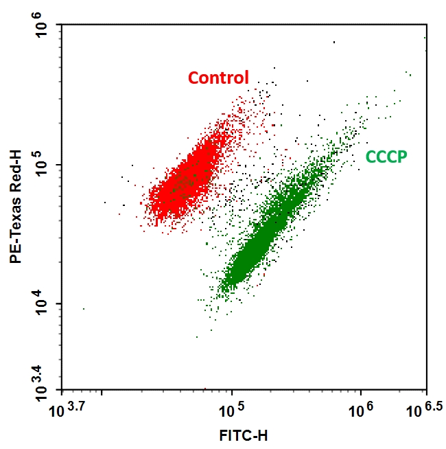
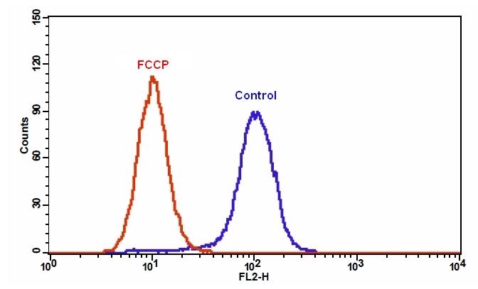
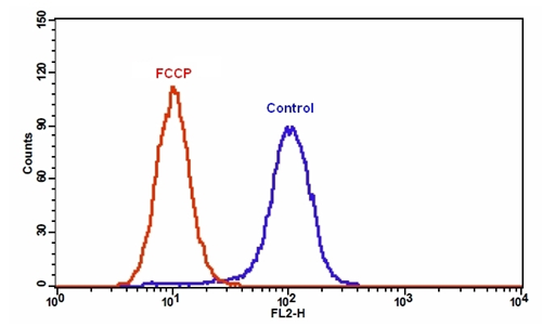
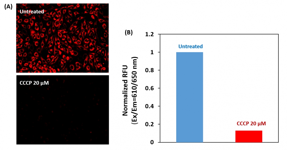
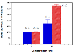
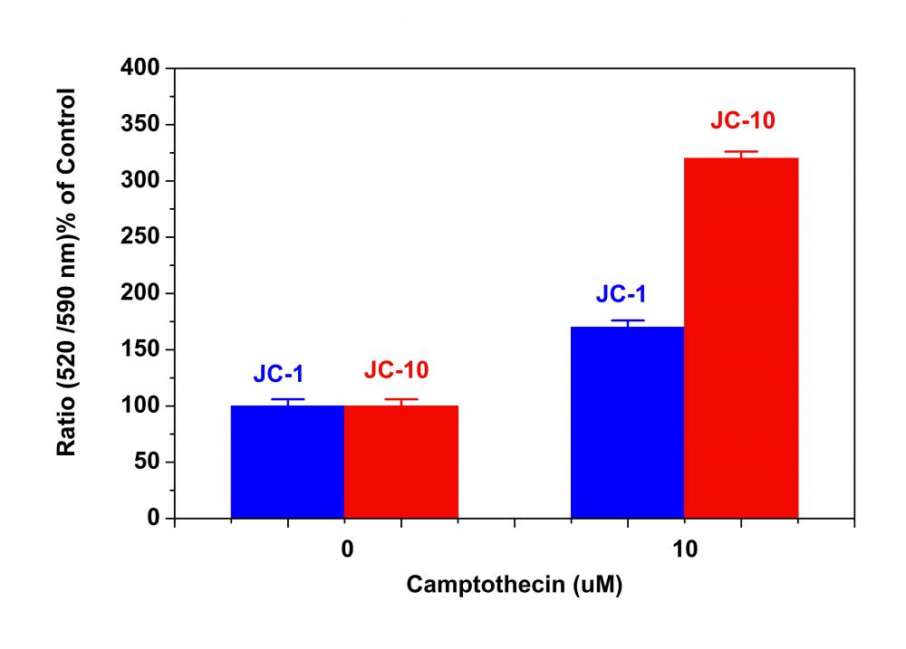
线粒体膜电位荧光探针JC-10是美国AAT Bioquest研发的用于检测线粒体膜电位的荧光探针是JC-1的完美替代品。JC-1在许多实验室中被广泛使用,但其水溶性差使得它在某些应用中难以使用。即使在1μM浓度下,JC-1也倾向于在水性缓冲液中沉淀。当需要高染料浓度时,JC-10已被开发为JC-1的替代物。与JC-1相比,我们的JC-10具有更好的水溶性。 JC-10能够选择性地进入线粒体,并随着膜电位的增加可逆地将其颜色从绿色变为橙色。这种性质是由于膜极化时JC-10聚集体的可逆形成导致发射光从520nm(即JC-10单体形式的发射)转变为570nm(即J-聚集体形式的发射)。当在490nm激发时,随着线粒体膜变得更加极化,JC-10的颜色从绿色橙色可逆地变为绿色橙色。使用通常安装在所有流式细胞仪中的过滤器可以检测两种颜色。可以在荧光通道1(FL1)中分析绿色发射,在通道2(FL2)中分析绿色橙色发射。除了用于流式细胞仪外,JC-10还可用于荧光成像。我们已经开发出一种在荧光微孔板平台中使用JC-10的方案。在一些细胞系中,JC-10具有优于JC-1的性能。金畔生物是AAT Bioquest 的中国代理商,为您提供最优质的线粒体膜电位荧光探针。

点击查看光谱

产品说明书

JC-10的分析方案

概述

准备含有测试化合物的细胞

添加JC-10工作溶液(100μL/孔用于96孔板或25μL/孔用于384孔板)

在室温或37 ℃孵育1小时

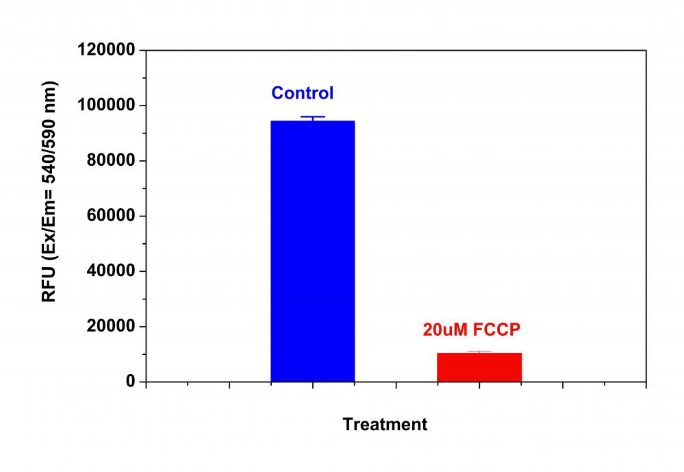
在Ex读取荧光强度 / Em = 490 / 525nm和540 / 590nm

注意:以下是我们推荐的活细胞方案。 该协议仅提供指南,应根据您的特定需求进行修改。


操作步骤

1.准备JC-10工作溶液:

1.1每瓶DMSO原液(100μL,2 mg / mL,3 mM)只能使用一次。任何未使用的小瓶应储存在<-20℃。注意:避免反复冻融循环,并避光.

1.2准备1X JC-10工作溶液:在实验当天,解冻一份JC-10 stockolution到室温。在Hanks和20 mM Hepesbuffer(HHBS)或您选择的缓冲液(pH 7-8,含0.02%Pluronic [表情] F-127)中制备10至30μM1X工作溶液。通过votexing将它们混合均匀。注意:对于某些细胞系,pH值为8的工作溶液可能会阻止JC-10泄漏。

2.用荧光酶标仪进行JC-10检测:

2.1用测试化合物处理细胞一段所需的时间(例如,Jurkat细胞可以用喜树碱处理4-6小时)以诱导细胞凋亡。 对于空白孔(没有细胞的培养基),加入相应量的化合物缓冲液。

2.2将100μL/孔/ 96孔板或25μL/孔/ 384孔板的JC-10工作溶液(来自步骤1.2)加入到细胞板中。

2.3将JC-10加载板在37 oC,5%CO2培养箱中孵育15-60分钟。

注意:适当的孵育时间取决于所使用的单个细胞类型和细胞浓度。优化每个实验的孵育时间。

2.4监测Ex / Em = 490/525 nm(FITC通道)和540/590 nm(TRITC通道)的荧光变化,进行比率分析。

可选:从板上取下JC-10工作溶液; 在分析之前,将100μL/孔/ 96孔板或25μL/孔/ 384孔HHBS板加回到细胞板中。

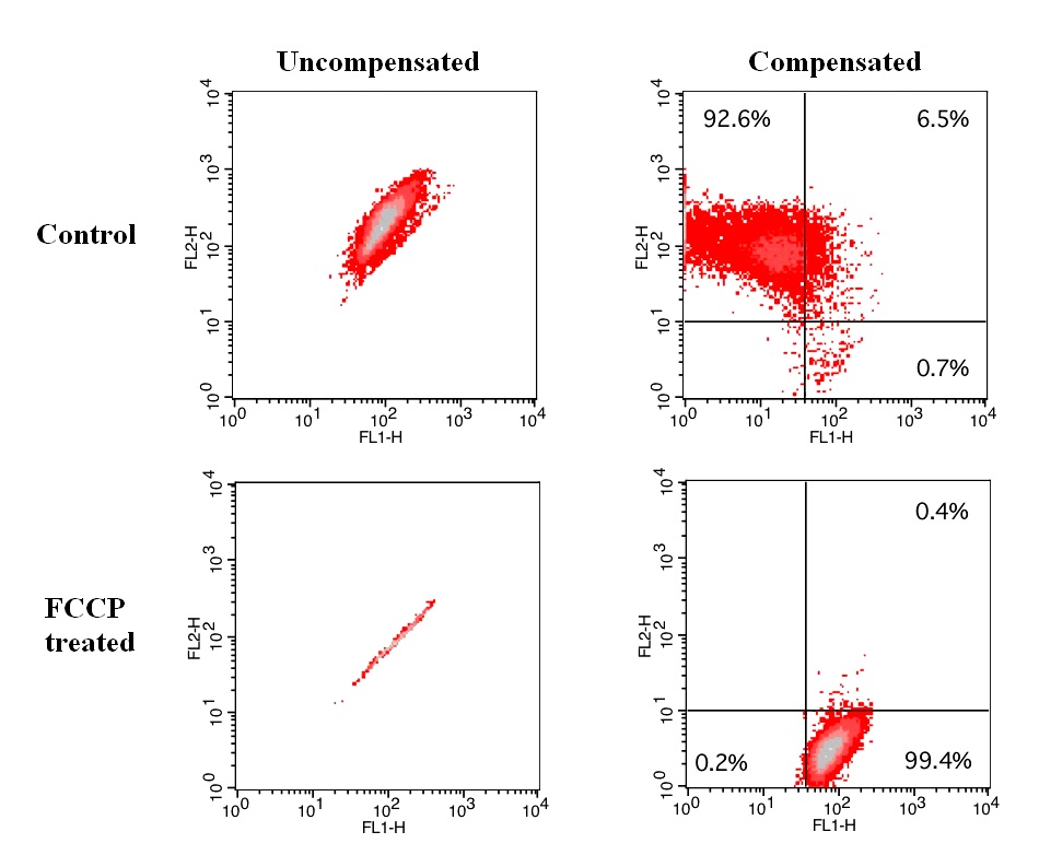
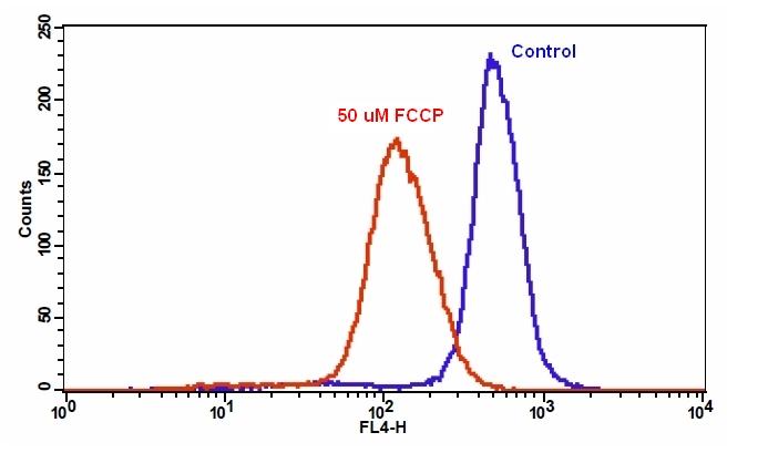
3.用荧光显微镜或流式细胞仪进行JC-10检测:

3.1用测试化合物处理细胞一段所需的时间(例如,Jurkat细胞可以用喜树碱处理4-6小时)以诱导细胞凋亡。

3.2离心细胞,每管取1-5×10⁵个细胞。

3.3将细胞重悬于500μLJC-10工作溶液中(来自步骤1.2)。

3.4在室温或37°C,5%CO2培养箱中孵育10至30分钟,避光。

注意:适当的孵育时间取决于所使用的单个细胞类型和细胞浓度。优化每个实验的孵育时间。

3.5使用荧光显微镜(使用FITC和TRITC过滤器)或流式细胞仪(使用FL1和FL2通道)监测Ex / Em = 490/525 nm和540/590 nm处的荧光变化。可选:移除JC-10工作 从板上解决; 在荧光显微镜下分析之前,将100μL/孔/ 96孔板或25μL/孔/ 384孔HHBS板加回到细胞板中。


参考文献

JC-10™ has been used to study many biologically significant processes across several key disciplines. To list a few, JC-10™ has been used to investigate topics such as mitochondrial membrane potential, cytotoxicity, cell viability, oxidative stress, cancer metastasis, apoptosis, signal transduction, mitochondrial fission and induced pluripotent stem cells (iPSCs). 

Below, you may find a small sampling of specific JC-10™ applications. To inquire about a potential application of JC-10™, or to consult with our fluorescent dye specialists, please contact us at support@aatbio.com or 1-800-990-8053.

High-content assays for hepatotoxicity using induced pluripotent stem cell–derived cells.
Researchers use JC-10™ to monitor mitochondrial depolarization as an indication of hepatotoxicity and oxidative stress in induced pluripotent stem cell-derived cells, with the ultimate goal of designing a reliable, high-content and imaging-based in vitro toxicity assay. 

High-Content High-Throughput Assays for Characterizing the Viability and Morphology of Human iPSC-Derived Neuronal Cultures
JC-10™ was used to study neurons derived from induced pluripotent stem-cells. Since JC-10™ will accumulate in the mitochondria of viable cells, it was used to determine cell viability in a high-throughput assay context.

Anticancer Activity of New Synthetic α-Methylene-δ-Lactones on Two Breast Cancer Cell Lines
Researchers chose JC-10™ to investigate mitochondrial membrane potential and membrane integrity in cells treated with natural products, such as α-Methylene-δ-Lactones, with the goal of developing new treatments for breast cancer.

Tetrandrine protects mouse retinal ganglion cells from ischemic injury.
JC-10™ was used in flow cytometry to study mitochondrial membrane potential (ΔΨm) in primary cultured retinal ganglion cells, as an extension of the field of drug discovery into prevention of ischemic injury. 

Midazolam induces cellular apoptosis in human cancer cells and inhibits tumor growth in xenograft mice
In a study of human cancer cells, JC-10™ was employed to track cellular apoptosis as a function of mitochondrial membrane potential, and consequently, mitochondrial activity. Researchers were interested in the possible anesthetic properties of midazolam for anticancer drug delivery.

Mitochondrial proteomics with siRNA knockdown to reveal ACAT1 and MDH2 in the development of doxorubicin-resistant uterine cancer
JC-10™ was used by researchers for the purposes of drug discovery. In particular, researchers were interested in finding new treatments for doxorubicin-resistant uterine cancer, using JC-10™ to monitor mitochondrial membrane potential and validate cell viability results. 

Cold exposure lowers energy expenditure at the cellular level
Researchers used JC-10™ to investigate the relationship between temperature and cellular activity. In particular, researchers wanted to explore if cold temperature acts as a stressor on mitochondrial membrane potential, with regards to the oxidative phosphorylation process which generates ATP.

Susceptibility of gametes and embryos of the eastern oyster, Crassostrea virginica, to Karenia brevis and its toxins
JC-10™ was used in the study of sperm viability, fertilization successs and embryonic survival of Crassostrea virginica. Specifically, JC-10™ was used to quantify mitochondrial membrane potential in sperm cells and to determine possible toxicity effects of algal blooms.

Calmodulin antagonists induce cell cycle arrest and apoptosis in vitro and inhibit tumor growth in vivo in human multiple myeloma
JC-10™ was used by researchers to study cell cycle and apoptosis in human multiple myeloma. JC-10™ functioned as a probe for the detection of mitochondrial membrane potential depolarization, which was crucial to the study of caspase activated apoptosis.

Activation of the mitochondrial apoptotic pathway produces reactive oxygen species and oxidative damage in hepatocytes that contribute to liver tumorigenesis
Researchers were interested in the pathways involved with liver tumorigenesis. To that end, they used JC-10™ to study activation of apoptotic pathways related to changes in mitochondrial membrane potential, with the ultimate goal of discovering if antioxidant therapy might help suppress liver carinogenesis.

说明书
线粒体膜电位荧光探针JC-10 JC-1的卓越代替品.pdf

MycoLight 比率细菌膜电位试剂盒 红色/绿色荧光 货号22401-AAT Bioquest荧光染料

上海金畔生物科技有限公司代理AAT Bioquest荧光染料全线产品,欢迎访问AAT Bioquest荧光染料官网了解更多信息。

MycoLight 比率细菌膜电位试剂盒 红色/绿色荧光

MycoLight 比率细菌膜电位试剂盒 红色/绿色荧光

MycoLight 比率细菌膜电位试剂盒 红色/绿色荧光     货号22401 货号 22401 存储条件 在零下15度以下保存, 避免光照
规格 200 Tests 价格 3924
Ex (nm) 483 Em (nm) 501
分子量 溶剂
产品详细介绍

简要概述

MycoLight 比率细菌膜电位试剂盒是美国AAT Bioquest生产的用于检测细菌的试剂盒,AAT Bioquest的MycoLight 比率细菌膜电位试剂盒使用荧光传感器,在低浓度的革兰氏阳性和阴性细菌细胞中均呈现绿色荧光,但由于较大的染料分子聚集,荧光在较高的细胞溶质浓度下向红色发射转移膜电位。 膜电位的大小随着不同的细菌种类而变化。 对于许多革兰氏阳性物种,红/绿比率倾向于随质子梯度的强度而变化,而在许多革兰氏阴性细菌中,染料的响应似乎与质子梯度强度不成比例。 该试剂盒设计用于在细菌浓度范围为每毫升105-107个生物体时测定细菌膜电位。 可以在510-530nm(FITC滤波片组)和600-660nm(德克萨斯红色滤波片组)下荧光测量染色细胞,激发波长为最常见的激发光源488nm。金畔生物是AAT Bioquest的中国代理商,为您提供最优质的MycoLight 比率细菌膜电位试剂盒。 

点击查看光谱


适用仪器


流式细胞仪  
激发: 488nm激光
发射: 530/30nm,610/20nm滤波片
推荐孔板: FITC,PE-Texas Red通道
荧光显微镜  
激发: 510/600nm
发射: 530/660nm
推荐孔板: 黑色透明
通道: FITC/Texas Red滤波片

产品说明书

操作方法

1.在任何适当的培养基中培养细菌从对数期培养物中获得健康细菌的最佳结果。在PBS(组分C)或等效的无菌缓冲液中将细菌培养物稀释至~106个细胞/ mL。可以直接从培养基中稀释细菌而不洗涤。准备足够的悬浮液,每次测试提供500 mL。

2.将500μl细菌悬浮液等分到流式细胞仪管中,进行每个染色实验。准备两个额外的管用于去极化控制和未染色的对照。

3.向去极化的对照样品中加入10μl500μMCCCP(组分B)并混合。

4.向每个流式细胞仪管中加入5μlMycoStainIt Green(100X)(组分A)并混合(不要在未染色的对照样品上加入染色剂)。在室温下孵育样品30分钟。可在5分钟后分析染色样品,但信号强度继续增加直至约30分钟。

5.可以在配备有发射488nm的激光的流式细胞仪中测定染色细菌。在绿色(荧光素滤光片)和红色(德克萨斯红色滤光片)通道中收集荧光。应使用对数信号放大收集前向散射,侧向散射和荧光。

6.仪器调整对于检测细菌等相对较小的颗粒尤其重要。使用未染色的对照样品定位前向和侧向散射通道中的细菌群。使用侧向散射作为设置采集触发器的参数。

7.如上所述,在调节流式细胞仪后应用去极化的对照样品。使用正向散射和侧向散射对细菌进行门控并调整荧光光电倍增管电压,使得绿色和红色MFI值近似相等。不要设置补偿。

8.虽然红色和绿色荧光强度的相对量将随着细胞大小和聚集而变化,但红色与绿色荧光强度的比率可以用作膜电位的尺寸无关指示。还可以通过使用正向散射与侧向散射对细菌进行门控来处理数据,并使用红色与绿色荧光的点图分析门控群体,将MFI值报告为线性值,而不是通道。

9.在比率直方图上,在感兴趣的峰周围设置标记并记录平均比值。对于红色与绿色荧光的点图,设置感兴趣群体周围的区域并记录每个的红色和绿色平均荧光强度(MFI)值。为了评估数据,将红色种群MFI除以绿色种群MFI。

10.在流式细胞仪中,细菌仅根据其大小和染色能力进行鉴定。最好通过荧光显微镜检查每个样品,以确认检测到的颗粒确实是细菌。


数据分析

MycoLight 比率细菌膜电位试剂盒 红色/绿色荧光     货号22401

图1.将枯草芽孢杆菌培养至对数期并在PBS中稀释至1×10 6个细胞/ mL的浓度。 然后将细胞用5μMCCCP处理20分钟,并与1X MycoStainIt Green孵育30分钟,然后进行流式细胞术分析。


参考文献

Raman spectroscopic analysis of Lactobacillus rhamnosus GG in response to dehydration reveals DNA conformation changes
Authors: Myintzu Hlaing, M.; Wood, B.; McNaughton, D.; Ying, D.; Augustin, M. A.
Journal: J Biophotonics (2017): 589-597

Antibacterial and antigelatinolytic effects of Satureja hortensis L. essential oil on epithelial cells exposed to Fusobacterium nucleatum
Authors: Zeidan-Chulia, F.; Keskin, M.; Kononen, E.; Uitto, V. J.; Soderling, E.; Moreira, J. C.; Gursoy, U. K.
Journal: J Med Food (2015): 503-6

Inactivation of Cronobacter sakazakii in reconstituted infant formula by combination of thymoquinone and mild heat
Authors: Shi, C.; Jia, Z.; Chen, Y.; Yang, M.; Liu, X.; Sun, Y.; Zheng, Z.; Zhang, X.; Song, K.; Cui, L.; Baloch, A. B.; Xia, X.
Journal: J Appl Microbiol (2015): 1700-6

Fourier transform infra-red spectroscopy and flow cytometric assessment of the antibacterial mechanism of action of aqueous extract of garlic (Allium sativum) against selected probiotic Bifidobacterium strains
Authors: Booyens, J.; Thantsha, M. S.
Journal: BMC Complement Altern Med (2014): 289

Deposition and survival of Escherichia coli O157:H7 on clay minerals in a parallel plate flow system
Authors: Cai, P.; Huang, Q.; Walker, S. L.
Journal: Environ Sci Technol (2013): 1896-903

Observation of injured E. coli population resulting from the application of high-pressure throttling treatments
Authors: De Lamo-Castellvi, S.; Toledo, R.; Frank, J. F.
Journal: J Food Sci (2013): M582-6

Effect of air drying on bacterial viability: A multiparameter viability assessment
Authors: Nocker, A.; Fernandez, P. S.; Montijn, R.; Schuren, F.
Journal: J Microbiol Methods (2012): 86-95

patients and environment
Authors: Lindback, T.; Rottenberg, M. E.; Roche, S. M.; Rorvik, L. M., The ability to enter into an avirulent viable but non-culturable (VBNC) form is widespread among Listeria monocytogenes isolates from salmon
Journal: Vet Res (2010): 8

Behaviors of physiologically active bacteria in water environment and chlorine disinfection
Authors: Sawaya, K.; Kaneko, N.; Fukushi, K.; Yaguchi, J.
Journal: Water Sci Technol (2008): 1343-8

Long-term survival of Legionella pneumophila in the viable but nonculturable state after monochloramine treatment
Authors: Alleron, L.; Merlet, N.; Lacombe, C.; Frere, J.
Journal: Curr Microbiol (2008): 497-502

说明书
MycoLight 比率细菌膜电位试剂盒 红色/绿色荧光 .pdf

Cell Meter 线粒体膜电位检测试剂盒 橙色荧光 适合流式细胞检测 货号22804-AAT Bioquest荧光染料

上海金畔生物科技有限公司代理AAT Bioquest荧光染料全线产品,欢迎访问AAT Bioquest荧光染料官网了解更多信息。

Cell Meter 线粒体膜电位检测试剂盒 橙色荧光 适合流式细胞检测

Cell Meter 线粒体膜电位检测试剂盒 橙色荧光 适合流式细胞检测

Cell Meter 线粒体膜电位检测试剂盒 橙色荧光 适合流式细胞检测     货号22804 货号 22804 存储条件 在零下15度以下保存, 避免光照
规格 100 Tests 价格 2604
Ex (nm) 546 Em (nm) 575
分子量 溶剂
产品详细介绍

简要概述

Cell Meter 线粒体膜电位检测试剂盒是美国AAT Bioquest生产的用于线粒体膜电位检测的试剂盒,Cell Meter检测试剂盒是一类用于检测细胞功能的系列工具,包括细胞活性、细胞毒性、细胞凋亡、细胞膜电位以及细胞周期等方面的指标。每种检测方案均能提供不同荧光颜色的检测方案。这些高效的检测方案为从多角度研究细胞功能活动提供了一种十分有效的方法。Cell Meter线粒体膜电位检测试剂盒 *橙色荧光 适合流式细胞检测* 专门通过检测线粒体跨膜电势的减少来检测细胞凋亡。线粒体跨膜电势瓦解与线粒体渗透转运孔的开放相一致,导致细胞色素C释放到胞浆中,它是下游凋亡级联反应的触发器。该荧光实验方案利用我们独有的MitoLite Orange来检测凋亡细胞中线粒体跨膜电势的减少。在正常细胞中,当MitoLite Orange积聚于线粒体时,红色荧光增加。然而在凋亡细胞中,MitoLite Orange随着线粒体跨膜电位的塌陷而减少。被MitoLite Orange染色的细胞可以通过488n的激发光(FL2 通道)来观察。该试剂盒可以与其它Cell Meter检测试剂盒同时检测细胞的活性和凋亡,该试剂盒已被优化,可用于流式细胞法筛选凋亡激活剂或抑制剂。金畔生物是AAT Bioquest的中国代理商,为您提供最优质的Cell Meter 线粒体膜电位检测试剂盒。 

点击查看光谱


适用仪器


流式细胞仪  
激发: 488nm 或 532nm激发
发射: 575/26nm滤波片
通道: PE通道

产品说明书

样品实验方案

简要概述

1.用密度为5×10⁵到1×10⁶个细胞/ mL的测试化合物制备细胞
2.将2μL500XMitoTell Orange加入1 mL细胞溶液中
3.将细胞在37℃,5%CO2培养箱中孵育15-30分钟
4.将细胞沉淀,并将细胞重悬于1mL生长培养基中
5.使用具有FL2通道的流式细胞仪分析细胞


操作步骤

1.对于每个样品,将细胞制备在1 mL温热培养基或您选择的缓冲液中,密度为5×10⁵至1×10⁶个细胞/ mL。注意:应对每个细胞系进行单独评估,以确定诱导细胞凋亡的最佳细胞密度。

2.用测试化合物处理细胞一段所需的时间以诱导细胞凋亡,并建立平行对照实验。

3.对于阴性对照:仅用载体处理细胞。

4.对于阳性对照:使用FCCP或CCCP以5-50μM在37℃,5%CO2培养箱中处理细胞15至30分钟。注意:CCCP或FCCP可与MitoTell Orange同时添加。为了获得最佳结果,可能需要对每个单独的细胞系滴定CCCP或FCCP。

5.将2μL500XMitoTell 橙(组分A)加入处理过的细胞中。

6.将细胞在37℃,5%CO2培养箱中孵育15至30分钟。注意:对于粘附细胞,用0.5 mM EDTA轻轻提起细胞以保持细胞完整,并在用MitoTell Orange染料加载溶液孵育之前用含有血清的培养基洗涤细胞一次。

7.将细胞以1000rpm离心4分钟,然后将细胞重悬于1mL的分析缓冲液(组分B)或您选择的缓冲液中。

8.使用具有FL2通道(Ex / Em = 540 / 590nm)的流式细胞仪监测荧光强度。


数据分析

Cell Meter 线粒体膜电位检测试剂盒 橙色荧光 适合流式细胞检测     货号22804

图1.在Jurkat细胞中添加FCCP后MitoTell Orange的荧光强度降低。 将Jurkat细胞单独加载MitoTell Orange(蓝色)或在30μMFCCP(红色)存在下加载15分钟。 使用FL2通道,用FACSCalibur(Becton Dickinson,San Jose,CA)流式细胞仪测量MitoTell Orange的荧光强度。


参考文献

Safranine O as a fluorescent probe for mitochondrial membrane potential studied on the single particle level and in suspension
Authors: Perevoshchikova IV, Sorochkina AI, Zorov DB, Antonenko YN.
Journal: Biochemistry (Mosc) (2009): 663

Computer-assisted live cell analysis of mitochondrial membrane potential, morphology and calcium handling
Authors: Koopman WJ, Distelmaier F, Esseling JJ, Smeitink JA, Willems PH.
Journal: Methods (2008): 304

Determination of high mitochondrial membrane potential in spermatozoa loaded with the mitochondrial probe 5,5′,6,6′-tetrachloro-1,1′,3,3′-tetraethylbenzimidazolyl-carbocyanine iodide (JC-1) by using fluorescence-activated flow cytometry
Authors: Guthrie HD, Welch GR.
Journal: Methods Mol Biol (2008): 89

Effects of eprosartan on mitochondrial membrane potential and H2O2 levels in leucocytes in hypertension
Authors: Labios M, Martinez M, Gabriel F, Guiral V, Ruiz-Aja S, Beltran B, Munoz A.
Journal: J Hum Hypertens (2008): 493

How DASPMI reveals mitochondrial membrane potential: fluorescence decay kinetics and steady-state anisotropy in living cells
Authors: Ramadass R, Bereiter-Hahn J.
Journal: Biophys J (2008): 4068

Life cell quantification of mitochondrial membrane potential at the single organelle level
Authors: Distelmaier F, Koopman WJ, Testa ER, de Jong AS, Swarts HG, Mayatepek E, Smeitink JA, Willems PH.
Journal: Cytometry A (2008): 129

Mitochondrial membrane potential in axons increases with local nerve growth factor or semaphorin signaling
Authors: Verburg J, Hollenbeck PJ.
Journal: J Neurosci (2008): 8306

The mitochondrial membrane potential and Ca2+ oscillations in smooth muscle
Authors: Chalmers S, McCarron JG.
Journal: J Cell Sci (2008): 75

[Evaluation of sperm mitochondrial membrane potential by JC-1 fluorescent staining and flow cytometry]
Authors: Xia XY, Wu YM, Hou BS, Yang B, Pan LJ, Shi YC, Jin BF, Shao Y, Cui YX, Huang YF.
Journal: Zhonghua Nan Ke Xue (2008): 135

Cyclosporin A-induced oxidative stress is not the consequence of an increase in mitochondrial membrane potential
Authors: van der Toorn M, Kauffman HF, van der Deen M, Slebos DJ, Koeter GH, Gans RO, Bakker SJ.
Journal: Febs J (2007): 3003


相关产品

产品名称 货号
Cell Meter 线粒体膜电位检测试剂盒 红色荧光 适合流式细胞检测 Cat#22806
Cell Meter 线粒体膜电位检测试剂盒 橙色荧光 适合微孔板检测 Cat#22805
Cell Meter 线粒体膜电位检测试剂盒 红色荧光 适合微孔板检测 Cat#22807

说明书
Cell Meter 线粒体膜电位检测试剂盒 橙色荧光 适合流式细胞检测 .pdf

线粒体膜电位荧光探针10-壬基吖啶橙溴化物 CAS 75168-11-5 货号22205-AAT Bioquest荧光染料

上海金畔生物科技有限公司代理AAT Bioquest荧光染料全线产品,欢迎访问AAT Bioquest荧光染料官网了解更多信息。

线粒体膜电位荧光探针10-壬基吖啶橙溴化物 CAS 75168-11-5

线粒体膜电位荧光探针10-壬基吖啶橙溴化物 CAS 75168-11-5

线粒体膜电位荧光探针10-壬基吖啶橙溴化物 CAS 75168-11-5    货号22205 货号 22205 存储条件 在零下15度以下保存, 避免光照
规格 25 mg 价格 1272
Ex (nm) 496 Em (nm) 519
分子量 472.5 溶剂 DMSO
产品详细介绍

简要概述

产品基本信息

货号:22205

产品名称:线粒体膜电位荧光探针10-壬基吖啶橙溴化物

CAS:75168-11-5

规格:25mg

储存条件:-15℃避光防潮

保质期:12个月


产品物理化学光谱特性

分子量:472.5

溶剂:DMSO

激发波长(nm):495

发射波长(nm):519


产品介绍

线粒体膜电位荧光探针10-壬基吖啶橙溴化物是美国AAT Bioquest生产的细胞膜电位荧光探针。10-壬基吖啶橙(NAO)是吖啶橙的衍生物, 是一种用于完整细胞内线粒体特异性荧光标记物。与Rh123相比,细胞中NAO的积聚与质子动力驱动关系不大,但是与线粒体相关膜蛋白或者脂类发生作用相关。NAO的摄入不依赖于线粒体跨膜电势差,可用于检测线粒体。NAO可与心磷脂等带负电荷磷脂特异性结合, 其与线粒体膜的相互作用不依赖于线粒体的膜电位; 分析细胞凋亡时, 可用罗丹明123来检测线粒体的膜电位, 而用NAO来检测线粒体结构的完整性。NAO和罗丹明123(Rh123)是研究线粒体功能一对好的染料。金畔生物是AAT Bioquest的中国代理商,为您提供最优质的线粒体膜电位荧光探针10-壬基吖啶橙溴化物。 

点击查看光谱


参考文献

Spectral studies of N-nonyl acridine orange in anionic, cationic and neutral surfactants
Authors: Wiosetek-Reske AM, Wysocki S.
Journal: Spectrochim Acta A Mol Biomol Spectrosc (2006): 1118

Binding of 10-N-nonyl acridine orange to cardiolipin-deficient yeast cells: implications for assay of cardiolipin
Authors: Gohil VM, Gvozdenovic-Jeremic J, Schlame M, Greenberg ML.
Journal: Anal Biochem (2005): 350

Determination of dipole moment in the ground and excited state by experimental and theoretical methods of N-nonyl acridine orange
Authors: Wiosetek-Reske AM, Wysocki S, Bak GW.
Journal: Spectrochim Acta A Mol Biomol Spectrosc (2005): 1172

Use of the fluorescent dye 10-N-nonyl acridine orange in quantitative and location assays of cardiolipin: a study on different experimental models
Authors: Garcia Fernandez MI, Ceccarelli D, Muscatello U.
Journal: Anal Biochem (2004): 174

Intracellular distribution of the fluorescent dye nonyl acridine orange responds to the mitochondrial membrane potential: implications for assays of cardiolipin and mitochondrial mass
Authors: Jacobson J, Duchen MR, Heales SJ.
Journal: J Neurochem (2002): 224

Cardiolipin binds nonyl acridine orange by aggregating the dye at exposed hydrophobic domains on bilayer surfaces
Authors: Mileykovskaya E, Dowhan W, Birke RL, Zheng D, Lutterodt L, Haines TH.
Journal: FEBS Lett (2001): 187

Staining of mitochondrial membranes with 10-nonyl acridine orange, MitoFluor Green, and MitoTracker Green is affected by mitochondrial membrane potential altering drugs
Authors: Keij JF, Bell-Prince C, Steinkamp JA.
Journal: Cytometry (2000): 203

Visualization of phospholipid domains in Escherichia coli by using the cardiolipin-specific fluorescent dye 10-N-nonyl acridine orange
Authors: Mileykovskaya E, Dowhan W.
Journal: J Bacteriol (2000): 1172

Direct cardiolipin assay in yeast using the red fluorescence emission of 10-N-nonyl acridine orange
Authors: Gallet PF, Maftah A, Petit JM, Denis-Gay M, Julien R.
Journal: Eur J Biochem (1995): 113

10N-nonyl acridine orange interacts with cardiolipin and allows the quantification of this phospholipid in isolated mitochondria
Authors: Petit JM, Maftah A, Ratinaud MH, Julien R.
Journal: Eur J Biochem (1992): 267

说明书
线粒体膜电位荧光探针10-壬基吖啶橙溴化物 CAS 75168-11-5.pdf

Cell Meter 线粒体膜电位检测试剂盒 红色荧光 适合流式细胞检测 货号22806-AAT Bioquest荧光染料

上海金畔生物科技有限公司代理AAT Bioquest荧光染料全线产品,欢迎访问AAT Bioquest荧光染料官网了解更多信息。

Cell Meter 线粒体膜电位检测试剂盒 红色荧光 适合流式细胞检测

Cell Meter 线粒体膜电位检测试剂盒 红色荧光 适合流式细胞检测

Cell Meter 线粒体膜电位检测试剂盒 红色荧光 适合流式细胞检测     货号22806 货号 22806 存储条件 在零下15度以下保存, 避免光照
规格 100 tests 价格 2604
Ex (nm) 613 Em (nm) 631
分子量 溶剂
产品详细介绍

简要概述

我们的Cell Meter 检测试剂盒是一套用于检测细胞功能的工具。可以使用多种参数。该特定试剂盒旨在通过测量线粒体膜电位(MMP)的丢失来检测细胞凋亡。线粒体膜电位的凋亡与线粒体通透性过渡孔的开放相吻合,导致细胞色素C释放到细胞质中,进而触发凋亡级联反应中的其他下游事件。该荧光测定法使用我们专有的阳离子MitoLite red检测细胞中线粒体膜电位的变化。在正常细胞中,当线粒体中积累了MitoLite red时,红色荧光强度会增加。但是,在凋亡细胞中,MMP凋亡后,MitoLite red的荧光强度降低。可以对用MitoLite red染色的细胞进行荧光检测。我们的Cell Meter 红色线粒体膜电位测定试剂盒可通过优化的测定方法提供所有必需成分。该试剂盒可用于筛选凋亡激活剂和抑制剂。该试剂盒可以使用流式细胞仪在APC或Cy5通道上观察用MitoTell Red染色的细胞。该试剂盒经过优化,可通过流式细胞仪筛选凋亡激活剂和抑制剂。金畔生物是AAT Bioquest的中国代理商,为您提供最优质的Cell Meter 线粒体膜电位检测试剂盒。

点击查看光谱


适用仪器


流式细胞仪  
激发: 640nm激光
发射: 660/20nm滤波片
通道: APC通道

产品说明书

样品实验方案

简要概述

  1. 用5×10⁵至1×10⁶细胞/ mL的密度制备含测试化合物的细胞
  2. 在0.5 mL细胞溶液中加入1 µL 500X MitoTell Red
  3. 将细胞在37°C,5%CO2培养箱中孵育15-30分钟
  4. 沉淀细胞,然后将细胞重悬于0.5 mL检测缓冲液中
  5. 使用带有APC或Cy5通道的流式细胞仪分析细胞


实验步骤

1.对于每个样品,在0.5 mL温暖的培养基或自备的缓冲液中以5×10⁵至1×10⁶细胞/ mL的密度制备细胞。注意:应单独评估每种细胞系,以确定诱导凋亡的最佳细胞密度。

2.用测试化合物处理细胞一段时间,以诱导细胞凋亡,并建立平行对照实验。注意:我们在37ºC下用20µM CCCP处理Jurkat细胞15分钟,以改变线粒体膜电位。CCCP或FCCP可以与MitoTell Red同时添加。为了获得最佳结果,可能需要为每个单独的细胞系滴定CCCP或FCCP。

3.向处理过的细胞中加入1 µL 500X MitoTell Red(组分A)。

4.将细胞在37°C,5%CO2培养箱中孵育15至30分钟。注意:对于贴壁细胞,用0.5 mM EDTA轻轻提起细胞以保持细胞完整,并在与MitoTell Red孵育之前用含血清的培养基洗涤细胞一次。

5.以800 rpm的速度离心细胞4分钟,然后将细胞重悬于0.5 mL的测定缓冲液(组分B)或自备的缓冲液中。

6.使用流式细胞仪通过APC或Cy5通道检测荧光强度。


参考文献

Safranine O as a fluorescent probe for mitochondrial membrane potential studied on the single particle level and in suspension
Authors: Perevoshchikova IV, Sorochkina AI, Zorov DB, Antonenko YN.
Journal: Biochemistry (Mosc) (2009): 663

Computer-assisted live cell analysis of mitochondrial membrane potential, morphology and calcium handling
Authors: Koopman WJ, Distelmaier F, Esseling JJ, Smeitink JA, Willems PH.
Journal: Methods (2008): 304

Determination of high mitochondrial membrane potential in spermatozoa loaded with the mitochondrial probe 5,5′,6,6′-tetrachloro-1,1′,3,3′-tetraethylbenzimidazolyl-carbocyanine iodide (JC-1) by using fluorescence-activated flow cytometry
Authors: Guthrie HD, Welch GR.
Journal: Methods Mol Biol (2008): 89

Effects of eprosartan on mitochondrial membrane potential and H2O2 levels in leucocytes in hypertension
Authors: Labios M, Martinez M, Gabriel F, Guiral V, Ruiz-Aja S, Beltran B, Munoz A.
Journal: J Hum Hypertens (2008): 493

Evaluation of sperm mitochondrial membrane potential by JC-1 fluorescent staining and flow cytometry
Authors: Xia XY, Wu YM, Hou BS, Yang B, Pan LJ, Shi YC, Jin BF, Shao Y, Cui YX, Huang YF.
Journal: Zhonghua Nan Ke Xue (2008): 135

How DASPMI reveals mitochondrial membrane potential: fluorescence decay kinetics and steady-state anisotropy in living cells
Authors: Ramadass R, Bereiter-Hahn J.
Journal: Biophys J (2008): 4068

Life cell quantification of mitochondrial membrane potential at the single organelle level
Authors: Distelmaier F, Koopman WJ, Testa ER, de Jong AS, Swarts HG, Mayatepek E, Smeitink JA, Willems PH.
Journal: Cytometry A (2008): 129

Mitochondrial membrane potential in axons increases with local nerve growth factor or semaphorin signaling
Authors: Verburg J, Hollenbeck PJ.
Journal: J Neurosci (2008): 8306

The mitochondrial membrane potential and Ca2+ oscillations in smooth muscle
Authors: Chalmers S, McCarron JG.
Journal: J Cell Sci (2008): 75

Cyclosporin A-induced oxidative stress is not the consequence of an increase in mitochondrial membrane potential
Authors: van der Toorn M, Kauffman HF, van der Deen M, Slebos DJ, Koeter GH, Gans RO, Bakker SJ.
Journal: Febs J (2007): 3003

说明书
Cell Meter 线粒体膜电位检测试剂盒 红色荧光 适合流式细胞检测 .pdf

Cell Meter 线粒体膜电位检测试剂盒 红色荧光 适合微孔板检测 货号22807-AAT Bioquest荧光染料

上海金畔生物科技有限公司代理AAT Bioquest荧光染料全线产品,欢迎访问AAT Bioquest荧光染料官网了解更多信息。

Cell Meter 线粒体膜电位检测试剂盒 红色荧光 适合微孔板检测

Cell Meter 线粒体膜电位检测试剂盒 红色荧光 适合微孔板检测

Cell Meter 线粒体膜电位检测试剂盒 红色荧光 适合微孔板检测     货号22807 货号 22807 存储条件 在零下15度以下保存, 避免光照
规格 500 tests 价格 2604
Ex (nm) 613 Em (nm) 631
分子量 溶剂
产品详细介绍

简要概述

我们的Cell Meter 检测试剂盒是一套用于检测细胞生存力的工具。可以使用多种参数。该特定试剂盒旨在通过测量线粒体膜电位(MMP)的消失来检测细胞凋亡。 MMP的凋亡与线粒体通透性过渡孔的开放相吻合,导致细胞色素C释放到细胞质中,进而触发了细胞凋亡级联反应中的其他下游事件。我们的Cell Meter 线粒体膜电位测定试剂盒可通过优化的测定方法提供所有基本成分。荧光测定法使用我们专有的阳离子MitoTell Red检测MMP消失的细胞凋亡。在正常细胞中,当MitoTell Red堆积在线粒体中时,红色荧光强度会增加。但是,在凋亡细胞中,MMP凋亡后,MitoTell Red的荧光强度降低。 MitoTell Red染色的细胞可以通过荧光显微镜Cy5通道或荧光酶标仪读取。该试剂盒经过优化,可通过荧光酶标仪读取凋亡激活剂和抑制剂。而且该测定可以以方便的96孔和384孔荧光酶标仪进行检测,而无需清洗步骤。金畔生物是AAT Bioquest的中国代理商,为您提供最优质的Cell Meter 线粒体膜电位检测试剂盒。

点击查看光谱


适用仪器


荧光酶标仪  
激发: 610nm
发射: 650nm
cutoff: 630nm
推荐孔板: 黑色透明
读取模式: 底读模式

产品说明书

样品实验方案

简要概述

  1. 准备细胞
  2. 添加测试化合物
  3. 添加MitoTell Red工作溶液(100 µL /孔/ 96孔板或25 µL /孔/ 384孔板)
  4. 将板在5%CO2、37°C的培养箱中孵育30分钟
  5. 添加测定缓冲液B(50 µL /孔/ 96孔板或12.5 µL /孔/ 384孔板)
  6. 在Ex / Em = 610/650 nm(截止= 630 nm)或使用Cy5滤光片的荧光显微镜下检测荧光增加(底部读取模式)


溶液配制

将50 µL的200X MitoTell Red(组分A)添加到10 mL的测定缓冲液A(组分B)中,并充分混合以制成MitoTell Red工作溶液,避光。


实验步骤

1.用测试化合物处理细胞一段时间,以诱导细胞凋亡,并建立平行对照实验。注意:我们用20 µM CCCP处理HeLa细胞15分钟,以改变线粒体膜电位。CCCP或FCCP可以与MitoTell Red同时添加。为了获得最佳结果,可能需要为每个单独的细胞系滴定CCCP或FCCP。

2.将100 µL /孔/ 96孔板或25 µL /孔/ 384孔板的MitoTell Red工作溶液加入细胞板。

3.在避光的条件下,将板在37ºC下孵育15-30分钟。注意:适当的孵育时间取决于所用的单个细胞类型和细胞浓度。优化每个实验的孵育时间。

4.将50 µL /孔/ 96孔板或12.5 µL /孔/ 384孔板的测定缓冲液B(组分C)添加到细胞板中。注意:加载后请勿洗涤细胞。对于非贴壁细胞,建议在加入测定缓冲液B(组分C)后,以800 rpm离心细胞板2分钟,然后制动。

5.加入测定缓冲液B(组分C)后10到30分钟,用荧光酶标仪(Ext / Em = 610/650 nm(截止= 630 nm))检测荧光强度,或在荧光下用带Cy5滤镜的显微镜观察荧光信号。


参考文献

Safranine O as a fluorescent probe for mitochondrial membrane potential studied on the single particle level and in suspension
Authors: Perevoshchikova IV, Sorochkina AI, Zorov DB, Antonenko YN.
Journal: Biochemistry (Mosc) (2009): 663

Computer-assisted live cell analysis of mitochondrial membrane potential, morphology and calcium handling
Authors: Koopman WJ, Distelmaier F, Esseling JJ, Smeitink JA, Willems PH.
Journal: Methods (2008): 304

Determination of high mitochondrial membrane potential in spermatozoa loaded with the mitochondrial probe 5,5′,6,6′-tetrachloro-1,1′,3,3′-tetraethylbenzimidazolyl-carbocyanine iodide (JC-1) by using fluorescence-activated flow cytometry
Authors: Guthrie HD, Welch GR.
Journal: Methods Mol Biol (2008): 89

Effects of eprosartan on mitochondrial membrane potential and H2O2 levels in leucocytes in hypertension
Authors: Labios M, Martinez M, Gabriel F, Guiral V, Ruiz-Aja S, Beltran B, Munoz A.
Journal: J Hum Hypertens (2008): 493

Evaluation of sperm mitochondrial membrane potential by JC-1 fluorescent staining and flow cytometry
Authors: Xia XY, Wu YM, Hou BS, Yang B, Pan LJ, Shi YC, Jin BF, Shao Y, Cui YX, Huang YF.
Journal: Zhonghua Nan Ke Xue (2008): 135

How DASPMI reveals mitochondrial membrane potential: fluorescence decay kinetics and steady-state anisotropy in living cells
Authors: Ramadass R, Bereiter-Hahn J.
Journal: Biophys J (2008): 4068

Life cell quantification of mitochondrial membrane potential at the single organelle level
Authors: Distelmaier F, Koopman WJ, Testa ER, de Jong AS, Swarts HG, Mayatepek E, Smeitink JA, Willems PH.
Journal: Cytometry A (2008): 129

Mitochondrial membrane potential in axons increases with local nerve growth factor or semaphorin signaling
Authors: Verburg J, Hollenbeck PJ.
Journal: J Neurosci (2008): 8306

The mitochondrial membrane potential and Ca2+ oscillations in smooth muscle
Authors: Chalmers S, McCarron JG.
Journal: J Cell Sci (2008): 75

Cyclosporin A-induced oxidative stress is not the consequence of an increase in mitochondrial membrane potential
Authors: van der Toorn M, Kauffman HF, van der Deen M, Slebos DJ, Koeter GH, Gans RO, Bakker SJ.
Journal: Febs J (2007): 3003

说明书
Cell Meter 线粒体膜电位检测试剂盒 红色荧光 适合微孔板检测 .pdf

Cell Meter JC-10线粒体膜电位检测试剂盒 适合微孔板检测 货号22800-AAT Bioquest荧光染料

上海金畔生物科技有限公司代理AAT Bioquest荧光染料全线产品,欢迎访问AAT Bioquest荧光染料官网了解更多信息。

Cell Meter JC-10线粒体膜电位检测试剂盒 适合微孔板检测

Cell Meter JC-10线粒体膜电位检测试剂盒 适合微孔板检测

Cell Meter JC-10线粒体膜电位检测试剂盒 适合微孔板检测     货号22800 货号 22800 存储条件 在零下15度以下保存, 避免光照
规格 500 Tests 价格 3924
Ex (nm) 508 Em (nm) 524
分子量 溶剂
产品详细介绍

简要概述

Cell Meter JC-10线粒体膜电位检测试剂盒 适合微孔板检测 是美国AAT Bioquest研发的用于检测线粒体膜电位的试剂盒,JC-1在许多实验室中被广泛使用,但其水溶性差导致极大的不便。即使在1μM浓度下,JC-1也倾向于在水性缓冲液中沉淀。当需要高染料浓度时,JC-10被开发成为JC的优异替代品。与JC-1相比,JC-10具有更好的水溶性。 JC-10能够选择性地进入线粒体,并随着膜电位的增加可逆地将其颜色从绿色变为橙色。该性质是由于膜极化时JC-10聚集体的可逆形成导致发射光从520nm(即JC-10单体形式的发射)向570nm(即J-聚集体形式的发射)的移动。当在490nm处激发时,随着线粒体膜变得更加极化,JC-10的颜色从绿色橙色可逆地变为绿色橙色。使用通常安装在所有流式细胞仪中的过滤器可以检测两种颜色。可以在荧光通道1(FL1)中分析绿色发射,在通道2(FL2)中分析绿色橙色发射。除了用于流式细胞仪外,它还可用于荧光成像和荧光微孔板平台。这种Cell Meter 线粒体膜电位检测试剂盒使研究人员能够以酶标仪的形式运行JC-10检测,并为监测线粒体膜电位变化提供了最强大的检测方法。

这种Cell Meter JC-10线粒体膜电位检测试剂盒使研究人员能够以酶标仪的形式运行JC-10检测,并为监测线粒体膜电位变化提供了最可靠的检测方法。该试剂盒基于阳离子亲脂性JC-10染料检测细胞中线粒体膜电位的变化。在正常细胞中,JC-10浓缩在线粒体基质中,在那里形成红色荧光聚集体。然而,在凋亡和坏死细胞中,JC-10扩散出线粒体。它变成单体形式并以绿色荧光染色细胞。该试剂盒可用于筛选凋亡激活剂和抑制剂。可以以方便的96孔和384孔荧光微量滴定板形式进行测定。金畔生物是AAT Bioquest 的中国代理商,为您提供最优质的线粒体膜电位荧光探针。

点击查看光谱


适用仪器


荧光酶标仪  
激发: 490/540nm
发射: 525/590nm
cutoff: 515/570nm
推荐孔板: 黑色透明
读取模式: 底读模式

产品说明书

96孔板样品分析

概述

准备细胞

添加测试化合物

添加JC-10染料加载溶液(50μL/孔/ 96孔板或12.5μL/孔/ 384孔板)

在37℃,5%CO2培养箱中孵育30至60分钟

添加测定 缓冲液B(50μL/孔/ 96孔板或12.5μL/孔/ 384孔板)

在Ex / Em = 490/525和540/590 nm处监测荧光强度


操作步骤

1.准备细胞:
1.1对于贴壁细胞:在生长培养基中将细胞以20,000至80,000个细胞/孔/90μL过夜,96孔板或5,000至20,000个细胞/孔/20μL,用于384孔板。
1.2对于非粘附细胞:从培养基中离心细胞,然后将细胞沉淀悬浮于100,000-200,000细胞/孔/90μL的培养基中,用于96孔poly-D赖氨酸平板或25,000-50,000细胞/孔 /20μL,用于384孔聚-D赖氨酸板。 在实验之前,在制动器关闭的情况下以800rpm离心板2分钟。
注意:应对每个细胞系进行单独评估,以确定诱导细胞凋亡的最佳细胞密度。


2.准备JC-10染料加载溶液:
2.1使用前在室温下清洗所有套件组件。
2.2将50μL100XJC-10(组分A)加入5mL测定缓冲液A(组分B)中,充分混合。
注意:将未使用的100X JC-10(组分A)等分并保存在-20 oC。 避免反复冻/融循环。


3.运行JC-10测定:
3.1通过向所需缓冲液(例如PBS或HHBS)中加入10μL10X测试化合物(96孔板)或5μL5X测试化合物(384-板)来处理细胞。
注意:在添加化合物之前不必洗涤细胞。然而,如果测试的化合物对血清敏感,则可在添加化合物之前吸出生长培养基和血清因子。在抽吸后将相同体积的HHBS添加到孔中(例如对于96孔板为90μL或对于384孔板为20μL)。或者,细胞可以在无血清培养基中生长。
3.2在室温或37℃,5%CO2培养箱中培养细胞板至少15分钟或所需的时间(对于Jurkat细胞,用喜树碱4-6小时或用星形孢菌素处理3-5小时)到诱导细胞凋亡
3.3将50μL/孔(96孔板)或12.5μL/孔(384孔板)的JC-10染料加载溶液(来自步骤2.2)加入细胞板(来自步骤3.2)。
3.4将染料加载板在37℃,5%CO2培养箱中孵育30-60分钟,避光。
注意:适当的孵育时间取决于所使用的单个细胞类型和细胞浓度。优化每个实验的孵育时间。
3.5在读取荧光强度之前,将50μL/孔(96孔板)或12.5μL/孔(384孔板)的测定缓冲液B(组分C)加入染料加载板(来自步骤3.4)。
注1:装载后请勿清洗电池。
注2:对于非粘附细胞,建议在加入分析缓冲液B(组分C)后,以800rpm离心细胞板2分钟,同时停止制动。
3.6使用底部读取模式监测Ex / Em = 490 / 525nm(在515nm处截止)和540 / 590nm(在570nm处截止)的荧光强度以进行比率分析。


数据分析

Em在525/590处的荧光强度比率用于数据分析。

Cell Meter JC-10线粒体膜电位检测试剂盒 适合微孔板检测     货号22800

图1.在Jurkat细胞中用JC-10和JC-1测量了喜树碱诱导的线粒体膜电位变化。 用喜树碱(10mM)处理Jurkat细胞4小时后,向孔中加入JC-1和JC-10染料上样溶液并孵育30分钟。 使用NOVOstar酶标仪(BMG Labtech)在Ex / Em = 490 / 525nm和490 / 590nm下测量J-聚集体和单体形式的JC-1和JC-10的荧光强度。


参考文献

Determination of Hepatotoxicity in iPSC-Derived Hepatocytes by Multiplexed High Content Assays
Authors: Oksana Sirenko, Evan F Cromwell
Journal: (2018): 339–354

Inhibitors of Aβ42-induced endoplasmic reticular unfolded protein response (UPRER), in yeast, also rescue yeast cells from Aβ42-mediated apoptosis
Authors: Asma Derf, Ramesh Mudududdla, Sandip B Bharate, Bhabatosh Chaudhuri
Journal: European Journal of Pharmaceutical Sciences (2018)

Knockout of Mpv17-Like Protein (M-LPH) Gene in Human Hepatoma Cells Results in Impairment of mtDNA Integrity through Reduction of TFAM, OGG1, and LIG3 at the Protein Levels
Authors: Reiko Iida, Misuzu Ueki, Toshihiro Yasuda
Journal: Oxidative Medicine and Cellular Longevity (2018)

Role of Mcl-1 in regulation of cell death in human induced pluripotent stem cell-derived cardiomyocytes in vitro
Authors: Liang Guo, Sandy Eldridge, Michael Furniss, Jodie Mussio, Myrtle Davis
Journal: Toxicology and Applied Pharmacology (2018)

Boosted Membrane Potential as Bioenergetic Response to Anoxia in Dinoroseobacter shibae
Authors: Christian Kirchhoff, Heribert Cypionka
Journal: Frontiers in Microbiology (2017): 695

Evaluation of anticancer properties of a new α-methylene-δ-lactone DL-249 on two cancer cell lines
Authors: Dorota K Pomorska, Katarzyna Gach-Janczak, Rafal Jakubowski, Tomasz Janecki, Jacek Szymański, Anna Janecka
Journal: Open Life Sciences (2017): 178–189

In vitro cardiotoxicity assessment of environmental chemicals using an organotypic human induced pluripotent stem cell-derived model
Authors: Oksana Sirenko, Fabian A Grimm, Kristen R Ryan, Yasuhiro Iwata, Weihsueh A Chiu, Frederick Parham, Jessica A Wignall, Blake Anson, Evan F Cromwell, Mamta Behl
Journal: Toxicology and Applied Pharmacology (2017)

Study of the relationship between AGEs and oxidative stress damage to trophoblast cell mitochondria
Authors: Lingling Jiang, Jianying Yan, Lixiang Wu
Journal: Ginekologia Polska (2017): 372–378

13-Methyl-palmatrubine induces apoptosis and cell cycle arrest in A549 cells in vitro and in vivo
Authors: Jingxian Chen, Xingang Lu, Chenghua Lu, Chunying Wang, Haizhu Xu, Xiaoli Xu, Haixin Gou, Bing Zhu, Wangchun Du
Journal: Oncology Reports (2016): 2526–2534

Anti-proliferation effects, efficacy of cyasterone in vitro and in vivo and its mechanism
Authors: XinGang Lu, HongFu Qiu, Liu Yang, JieYing Zhang, ShuJie Ma, Lan Zhen
Journal: Biomedicine & Pharmacotherapy (2016): 330–339


相关产品

产品名称 货号
Cell Meter JC-10线粒体膜电位检测试剂盒 适合流式细胞检测 Cat#22801

说明书
Cell Meter JC-10线粒体膜电位检测试剂盒 适合微孔板检测 .pdf

Cell Meter JC-10线粒体膜电位检测试剂盒 适合流式细胞检测 货号22801-AAT Bioquest荧光染料

上海金畔生物科技有限公司代理AAT Bioquest荧光染料全线产品,欢迎访问AAT Bioquest荧光染料官网了解更多信息。

Cell Meter JC-10线粒体膜电位检测试剂盒 适合流式细胞检测

Cell Meter JC-10线粒体膜电位检测试剂盒 适合流式细胞检测

Cell Meter JC-10线粒体膜电位检测试剂盒 适合流式细胞检测     货号22801 货号 22801 存储条件 在零下15度以下保存, 避免光照
规格 100 Tests 价格 3924
Ex (nm) 508 Em (nm) 524
分子量 溶剂
产品详细介绍

简要概述

Cell Meter JC-10线粒体膜电位检测试剂盒 适合流式细胞仪检测 是美国AAT Bioquest研发的用于检测线粒体膜电位的试剂盒,JC-1在许多实验室中被广泛使用,但其水溶性差导致极大的不便。即使在1μM浓度下,JC-1也倾向于在水性缓冲液中沉淀。当需要高染料浓度时,JC-10被开发成为JC的优异替代品。与JC-1相比,JC-10具有更好的水溶性。 JC-10能够选择性地进入线粒体,并随着膜电位的增加可逆地将其颜色从绿色变为橙色。该性质是由于膜极化时JC-10聚集体的可逆形成导致发射光从520nm(即JC-10单体形式的发射)向570nm(即J-聚集体形式的发射)的移动。当在490nm处激发时,随着线粒体膜变得更加极化,JC-10的颜色从绿色橙色可逆地变为绿色橙色。使用通常安装在所有流式细胞仪中的过滤器可以检测两种颜色。可以在荧光通道1(FL1)中分析绿色发射,在通道2(FL2)中分析绿色橙色发射。除了用于流式细胞仪外,它还可用于荧光成像和荧光微孔板平台。这种Cell Meter 线粒体膜电位检测试剂盒使研究人员能够以酶标仪的形式运行JC-10检测,并为监测线粒体膜电位变化提供了最强大的检测方法。

这种Cell Meter JC-10线粒体膜电位检测试剂盒使研究人员能够以酶标仪的形式运行JC-10检测,并为监测线粒体膜电位变化提供了最可靠的检测方法。该试剂盒基于阳离子亲脂性JC-10染料检测细胞中线粒体膜电位的变化。在正常细胞中,JC-10浓缩在线粒体基质中,在那里形成红色荧光聚集体。然而,在凋亡和坏死细胞中,JC-10扩散出线粒体。它变成单体形式并以绿色荧光染色细胞。该试剂盒可用于筛选凋亡激活剂和抑制剂。可以以方便的96孔和384孔荧光微量滴定板形式进行测定。金畔生物是AAT Bioquest 的中国代理商,为您提供最优质的线粒体膜电位荧光探针。

点击查看光谱


适用仪器


流式细胞仪  
激发: 488nm激光
发射: 530/30 nm, 575/26 nm滤波片
通道: FITC,PE通道

产品说明书

96孔板样品分析

概述

用密度为5×10⁵到1×10⁶个细胞/ mL的测试化合物制备细胞

用500μL1XJC-10染料加载溶液重悬细胞(2-5×10⁵细胞/管)

37°C孵育/ 室温15-30分钟

用流式细胞仪分析FL1通道(绿色荧光单体信号)和FL2通道(橙色荧光聚合信号)


操作方法

1.准备细胞:
以5×10⁵至1×10⁶个细胞/ mL的密度制备细胞。
注意:应根据个体评估每个细胞系,以确定诱导细胞凋亡的最佳细胞密度。


2.准备JC-10染料加载溶液:
2.1使用前在室温下清洗所有试剂盒中的试剂。
2.2将25μL的200X JC-10(组分A)加入5mL的测定缓冲液(组分B)中,并充分混合。
注意:将未使用的200X JC-10(组分A)等分并保存在-20℃。 避免反复冻/融循环。


3.运行JC-10测定:
3.1用测试化合物细胞培养所需的一段时间以诱导细胞凋亡。设置并行控制实验。
对于阴性对照:仅用载体处理细胞。
对于阳性对照:在37℃,5%CO2培养箱中用2-10μM的FCCP或CCCP处理细胞15至30分钟。
注意:CCCP或FCCP可与JC-10染料加载溶液同时添加(来自步骤2.2)。可能需要滴定CCCP或FCCP以获得具有单个细胞系的最佳结果。
3.2离心细胞,每管2-5×10⁵个细胞。
注意:对于贴壁细胞,用0.5 mM EDTA轻轻提起细胞以保持细胞摄入,用含有血清的培养基洗涤细胞一次,然后用JC-10染料加载溶液孵育(参见步骤3.3)。
3.3在500μLJC-10染料加载溶液中重悬细胞(来自步骤2.2)。
3.4在室温下或在37℃,5%CO2培养箱中孵育染料装载细胞15-60分钟,避光。
注意:适当的孵育时间取决于所使用的单个细胞类型和细胞浓度。优化每个实验的孵育时间。
3.5通过在FL1通道中使用流式细胞仪检测荧光强度,获得绿色荧光单体信号(在凋亡细胞中),并使用FL2通道检测橙色荧光聚集信号(在健康细胞中)。建议使用FCCP或CCCP处理的细胞进行补偿校正(见附录)。


数据分析

1.在活的非凋亡细胞中,JC-10作为细胞溶质染色绿色中的单体存在,并且还在线粒体染成红色的聚集体中积累。 在凋亡细胞和死细胞中,JC-10仅以单体形式存在,染色细胞溶质绿。
2.使用FL2通道可检测到健康细胞中含有红色JC-10聚集体的线粒体。 通过使用FL1通道(FITC)可检测凋亡细胞中的绿色JC-10单体。
3. FL1与FL2的强度比用于监测化合物处理诱导的线粒体膜电位变化。

Cell Meter JC-10线粒体膜电位检测试剂盒 适合流式细胞检测     货号22801

图1. FCCP诱导的JurKat细胞线粒体膜电位变化的影响。 将JurKat细胞染色载有JC-10染料加载溶液以及DMSO(Top)或5μMFCCP(低)10分钟。 使用FL1和FL2通道,用FACSCalibur(Becton Dickinson,San Jose,CA)流式细胞仪测量J-聚集体和单体形式JC-10的荧光强度。 将未补偿的数据(左栏)与补偿数据(右栏)进行比较。


参考文献

Anticancer properties of a new non-oxido vanadium (IV) complex with a catechol-modified 3, 3′-diindolylmethane ligand
Authors: Katja Dankhoff, Aamir Ahmad, Birgit Weber, Bernhard Biersack, Rainer Schobert
Journal: Journal of Inorganic Biochemistry (2019)

BDE-47 decreases progesterone levels in BeWo cells by interfering with mitochondrial functions and genes related to cholesterol transport
Authors: Anqi Shan, Mengxue Li, Xuejun Li, Yaoyan Li, Mengfan Yan, Ping Xian, Ying Chang, Xi Chen, Nai-jun Tang
Journal: Chemical Research in Toxicology (2019)

Enhancing magnetic resonance/photoluminescence imaging-guided photodynamic therapy by multiple pathways
Authors: Pei Liu, Jinghua Ren, Yuxuan Xiong, Zhe Yang, Wei Zhu, Qianyuan He, Zushun Xu, Wenshan He, Jing Wang
Journal: Biomaterials (2019)

5-Fluorouracil inhibits neural differentiation via Mfn1/2 reduction in human induced pluripotent stem cells
Authors: Shigeru Yamada, Daiju Yamazaki, Yasunari Kanda
Journal: The Journal of Toxicological Sciences (2018): 727–734

Determination of Hepatotoxicity in iPSC-Derived Hepatocytes by Multiplexed High Content Assays
Authors: Oksana Sirenko, Evan F Cromwell
Journal: (2018): 339–354

Yap1 safeguards mouse embryonic stem cells from excessive apoptosis during differentiation
Authors: Lucy LeBlanc, Bum-Kyu Lee, C Yu Andy, Mijeong Kim, Aparna V Kambhampati, Shannon M Dupont, Davide Seruggia, Byoung U Ryu, Stuart H Orkin, Jonghwan Kim
Journal: eLife (2018): e40167

Boosted Membrane Potential as Bioenergetic Response to Anoxia in Dinoroseobacter shibae
Authors: Christian Kirchhoff, Heribert Cypionka
Journal: Frontiers in Microbiology (2017): 695

Evaluation of anticancer properties of a new α-methylene-δ-lactone DL-249 on two cancer cell lines
Authors: Dorota K Pomorska, Katarzyna Gach-Janczak, Rafal Jakubowski, Tomasz Janecki, Jacek Szymański, Anna Janecka
Journal: Open Life Sciences (2017): 178–189

In vitro cardiotoxicity assessment of environmental chemicals using an organotypic human induced pluripotent stem cell-derived model
Authors: Oksana Sirenko, Fabian A Grimm, Kristen R Ryan, Yasuhiro Iwata, Weihsueh A Chiu, Frederick Parham, Jessica A Wignall, Blake Anson, Evan F Cromwell, Mamta Behl
Journal: Toxicology and Applied Pharmacology (2017)

Study of the relationship between AGEs and oxidative stress damage to trophoblast cell mitochondria
Authors: Lingling Jiang, Jianying Yan, Lixiang Wu
Journal: Ginekologia Polska (2017): 372–378


相关产品

产品名称 货号
Cell Meter JC-10线粒体膜电位检测试剂盒 适合微孔板检测 Cat#22800

说明书
Cell Meter JC-10线粒体膜电位检测试剂盒 适合流式细胞检测 .pdf

Cell Meter 线粒体膜电位近红外检测试剂盒 适合流式细胞检测 货号22802-AAT Bioquest荧光染料

上海金畔生物科技有限公司代理AAT Bioquest荧光染料全线产品,欢迎访问AAT Bioquest荧光染料官网了解更多信息。

Cell Meter 线粒体膜电位近红外检测试剂盒 适合流式细胞检测

Cell Meter 线粒体膜电位近红外检测试剂盒 适合流式细胞检测

Cell Meter 线粒体膜电位近红外检测试剂盒 适合流式细胞检测     货号22802 货号 22802 存储条件 在零下15度以下保存, 避免光照
规格 100 Tests 价格 2604
Ex (nm) Em (nm)
分子量 溶剂
产品详细介绍

简要概述

我们的Cell Meter 检测试剂盒是一套用于检测细胞生存力的工具。有多种参数可用于检测细胞活力。该特定试剂盒旨在通过测量线粒体膜电位的丧失来检测细胞凋亡。线粒体膜电位的崩溃与线粒体通透性过渡孔的开放相吻合,导致细胞色素C释放到细胞质中,进而触发凋亡级联反应中的其他下游事件。我们的Cell Meter NIR膜电位检测试剂盒为所有必需成分提供了优化的测定方法,可检测细胞凋亡的线粒体膜电位。该荧光测定法是基于我们专有的阳离子MitoLite NIR 染料对细胞中线粒体膜电位的检测。在正常细胞中,MitoLite NIR 主要在线粒体中积累,但是在凋亡细胞中,MitoLite NIR 染色强度降低。通过MitoLite NIR 染色的细胞可以通过流式细胞术观察到红色激发和远红色发射(FL4通道)。该试剂盒可与其他试剂配对,例如蓝激发碘化丙啶和Cell Meter 磷脂酰丝氨酸凋亡检测试剂盒(#22803),用于多参数研究细胞活力和凋亡。该试剂盒经过优化,可通过流式细胞仪筛选凋亡激活剂和抑制剂。金畔生物是AAT Bioquest的中国代理商,为您提供最优质的Cell Meter 线粒体膜电位近红外检测试剂盒。

点击查看光谱


适用仪器


流式细胞仪  
激发: 640nm激光
发射: 660/20nm滤波片
通道: APC通道

产品说明书

样品实验方案

简要概述

  1. 用5×10⁵至1×10⁶细胞/ mL的密度制备含测试化合物的细胞
  2. 将5 µL 200X MitoLite NIR加入1 mL细胞溶液中
  3. 在37°C,5%CO2的培养箱中孵育细胞15-30分钟
  4. 沉淀细胞,并将细胞重悬于1 mL生长培养基中
  5. 使用带有FL4通道的流式细胞仪分析细胞


操作步骤

1.对于每个样品,在1 mL温暖的培养基或您选择的缓冲液中以5×10⁵至1×10⁶细胞/ mL的密度制备细胞。 注意:应单独评估每种细胞系,以确定诱导凋亡的最佳细胞密度。

2.用测试化合物处理细胞一段时间,以诱导细胞凋亡,并建立平行对照实验。

阴性对照:仅用载体处理细胞。

阳性对照:在37℃,5%CO2培养箱中,以5-50 µM的浓度用FCCP或CCCP处理细胞15至30分钟。 注意:CCCP或FCCP可以与MitoLite NIR同时添加。 为了获得最佳结果,可能需要为每个单独的细胞系滴定CCCP或FCCP。

3.向处理过的细胞中加入5 µL 200X MitoLite NIR(组分A)。

4.将细胞在37°C,5%CO2培养箱中孵育15至30分钟。 注意:对于贴壁细胞,在用MitoLite NIR染料上样溶液孵育之前,用0.5 mM EDTA轻轻提起细胞以保持细胞完整,并用含血清的培养基洗涤一次。

5.以1000 rpm离心细胞4分钟,然后将细胞重悬于1 mL分析缓冲液(组分B)或您选择的缓冲液中。

6.使用带有FL4通道的流式细胞仪(Ex / Em = 635/660 nm)检测荧光强度。


参考文献

Safranine O as a fluorescent probe for mitochondrial membrane potential studied on the single particle level and in suspension
Authors: Perevoshchikova IV, Sorochkina AI, Zorov DB, Antonenko YN.
Journal: Biochemistry (Mosc) (2009): 663

Computer-assisted live cell analysis of mitochondrial membrane potential, morphology and calcium handling
Authors: Koopman WJ, Distelmaier F, Esseling JJ, Smeitink JA, Willems PH.
Journal: Methods (2008): 304

Determination of high mitochondrial membrane potential in spermatozoa loaded with the mitochondrial probe 5,5′,6,6′-tetrachloro-1,1′,3,3′-tetraethylbenzimidazolyl-carbocyanine iodide (JC-1) by using fluorescence-activated flow cytometry
Authors: Guthrie HD, Welch GR.
Journal: Methods Mol Biol (2008): 89

Effects of eprosartan on mitochondrial membrane potential and H2O2 levels in leucocytes in hypertension
Authors: Labios M, Martinez M, Gabriel F, Guiral V, Ruiz-Aja S, Beltran B, Munoz A.
Journal: J Hum Hypertens (2008): 493

Evaluation of sperm mitochondrial membrane potential by JC-1 fluorescent staining and flow cytometry
Authors: Xia XY, Wu YM, Hou BS, Yang B, Pan LJ, Shi YC, Jin BF, Shao Y, Cui YX, Huang YF.
Journal: Zhonghua Nan Ke Xue (2008): 135

How DASPMI reveals mitochondrial membrane potential: fluorescence decay kinetics and steady-state anisotropy in living cells
Authors: Ramadass R, Bereiter-Hahn J.
Journal: Biophys J (2008): 4068

Life cell quantification of mitochondrial membrane potential at the single organelle level
Authors: Distelmaier F, Koopman WJ, Testa ER, de Jong AS, Swarts HG, Mayatepek E, Smeitink JA, Willems PH.
Journal: Cytometry A (2008): 129

Mitochondrial membrane potential in axons increases with local nerve growth factor or semaphorin signaling
Authors: Verburg J, Hollenbeck PJ.
Journal: J Neurosci (2008): 8306

The mitochondrial membrane potential and Ca2+ oscillations in smooth muscle
Authors: Chalmers S, McCarron JG.
Journal: J Cell Sci (2008): 75

Cyclosporin A-induced oxidative stress is not the consequence of an increase in mitochondrial membrane potential
Authors: van der Toorn M, Kauffman HF, van der Deen M, Slebos DJ, Koeter GH, Gans RO, Bakker SJ.
Journal: Febs J (2007): 3003

说明书
Cell Meter 线粒体膜电位近红外检测试剂盒 适合流式细胞检测 .pdf

Cell Meter 线粒体膜电位检测试剂盒 橙色荧光 适合微孔板检测 货号22805-AAT Bioquest荧光染料

上海金畔生物科技有限公司代理AAT Bioquest荧光染料全线产品,欢迎访问AAT Bioquest荧光染料官网了解更多信息。

Cell Meter 线粒体膜电位检测试剂盒 橙色荧光 适合微孔板检测

Cell Meter 线粒体膜电位检测试剂盒 橙色荧光 适合微孔板检测

Cell Meter 线粒体膜电位检测试剂盒 橙色荧光 适合微孔板检测     货号22805 货号 22805 存储条件 在零下15度以下保存, 避免光照
规格 500 Tests 价格 2604
Ex (nm) 546 Em (nm) 575
分子量 溶剂
产品详细介绍

简要概述

我们的Cell Meter 检测试剂盒是一套用于检测细胞功能的工具。可以使用多种参数。该特定试剂盒旨在通过测量线粒体膜电位(MMP)的丢失来检测细胞凋亡。线粒体膜电位的凋亡与线粒体通透性过渡孔的开放相吻合,导致细胞色素C释放到细胞质中,进而触发凋亡级联反应中的其他下游事件。该荧光测定法使用我们专有的阳离子MitoLite Orange检测细胞中线粒体膜电位的变化。在正常细胞中,当线粒体中积累了MitoLite Orange时,橙色荧光强度会增加。但是,在凋亡细胞中,MMP凋亡后,MitoLite Orange的荧光强度降低。可以对用MitoLite Orange染色的细胞进行荧光检测。我们的Cell Meter 橙色线粒体膜电位测定试剂盒可通过优化的测定方法提供所有必需成分。该试剂盒可用于筛选凋亡激活剂和抑制剂。而且该测定可以以方便的96孔和384孔荧光酶标仪进行检测,而无需清洗步骤。金畔生物是AAT Bioquest的中国代理商,为您提供最优质的Cell Meter 线粒体膜电位检测试剂盒。


适用仪器


荧光酶标仪  
激发: 540nm
发射: 590nm
cutoff: 570nm
推荐孔板: 黑色透明
读取模式: 底读模式

产品说明书

样品实验方案

简要概述

  1. 准备细胞
  2. 添加测试化合物
  3. 添加MitoTell Orange工作溶液(100 µL /孔/ 96孔板或25 µL /孔/ 384孔板)
  4. 将板在5%CO2、37°C的培养箱中孵育15-30分钟
  5. 添加测定缓冲液B(50 µL /孔/ 96孔板或12.5 µL /孔/ 384孔板)
  6. 检测Ex / Em = 540/590 nm(截止= 570 nm)的荧光增加(底部读取模式)


溶液配制

工作溶液配制

将50 µL的200X MitoTell 橙色(组分A)添加到10 mL的测定缓冲液A(组分B)中,并充分混合以制成MitoTell Orange工作溶液,避光。


实验步骤

1.用测试化合物处理细胞一段时间,以诱导细胞凋亡,并建立平行对照实验。

阴性对照:仅用载体处理细胞。

阳性对照:在37°C 5%CO2培养箱中,以5-50 µM的浓度用FCCP或CCCP处理细胞15至30分钟。 注意:CCCP或FCCP可以与MitoTell Orange同时添加。 为了获得最佳结果,可能需要为每个单独的细胞系滴定CCCP或FCCP。

2.取出细胞培养基。注意:在添加MitoTell Orange工作溶液之前,必须除去细胞培养基。

3.将100 µL /孔/ 96孔板或25 µL /孔/ 384孔板的MitoTell Orange工作溶液添加到细胞板中。

4.将板在5%CO2、37°C​​的培养箱中避光放置15-30分钟。注意:适当的孵育时间取决于所用的单个细胞类型和细胞浓度。优化每个实验的孵育时间。

5.将50 µL /孔/ 96孔板或12.5 µL /孔/ 384孔板的测定缓冲液B(组分C)添加到细胞板中。注意:加载后请勿洗涤细胞。对于非贴壁细胞,建议在加入测定缓冲液B(组分C)后,以800 rpm离心细胞板2分钟,然后制动。

6.在加入分析缓冲液B(成分C)10至30分钟后,使用荧光酶标仪(底部读取模式)在Ex/Em=640/680 nm(截止=665 nm)处检测荧光强度,可以使用终点模式,也可以使用动力学模式。


参考文献

Safranine O as a fluorescent probe for mitochondrial membrane potential studied on the single particle level and in suspension
Authors: Perevoshchikova IV, Sorochkina AI, Zorov DB, Antonenko YN.
Journal: Biochemistry (Mosc) (2009): 663

Computer-assisted live cell analysis of mitochondrial membrane potential, morphology and calcium handling
Authors: Koopman WJ, Distelmaier F, Esseling JJ, Smeitink JA, Willems PH.
Journal: Methods (2008): 304

Determination of high mitochondrial membrane potential in spermatozoa loaded with the mitochondrial probe 5,5′,6,6′-tetrachloro-1,1′,3,3′-tetraethylbenzimidazolyl-carbocyanine iodide (JC-1) by using fluorescence-activated flow cytometry
Authors: Guthrie HD, Welch GR.
Journal: Methods Mol Biol (2008): 89

Effects of eprosartan on mitochondrial membrane potential and H2O2 levels in leucocytes in hypertension
Authors: Labios M, Martinez M, Gabriel F, Guiral V, Ruiz-Aja S, Beltran B, Munoz A.
Journal: J Hum Hypertens (2008): 493

Evaluation of sperm mitochondrial membrane potential by JC-1 fluorescent staining and flow cytometry
Authors: Xia XY, Wu YM, Hou BS, Yang B, Pan LJ, Shi YC, Jin BF, Shao Y, Cui YX, Huang YF.
Journal: Zhonghua Nan Ke Xue (2008): 135

How DASPMI reveals mitochondrial membrane potential: fluorescence decay kinetics and steady-state anisotropy in living cells
Authors: Ramadass R, Bereiter-Hahn J.
Journal: Biophys J (2008): 4068

Life cell quantification of mitochondrial membrane potential at the single organelle level
Authors: Distelmaier F, Koopman WJ, Testa ER, de Jong AS, Swarts HG, Mayatepek E, Smeitink JA, Willems PH.
Journal: Cytometry A (2008): 129

Mitochondrial membrane potential in axons increases with local nerve growth factor or semaphorin signaling
Authors: Verburg J, Hollenbeck PJ.
Journal: J Neurosci (2008): 8306

The mitochondrial membrane potential and Ca2+ oscillations in smooth muscle
Authors: Chalmers S, McCarron JG.
Journal: J Cell Sci (2008): 75

Cyclosporin A-induced oxidative stress is not the consequence of an increase in mitochondrial membrane potential
Authors: van der Toorn M, Kauffman HF, van der Deen M, Slebos DJ, Koeter GH, Gans RO, Bakker SJ.
Journal: Febs J (2007): 3003

说明书
Cell Meter 线粒体膜电位检测试剂盒 橙色荧光 适合微孔板检测 .pdf

线粒体膜电位荧光探针罗丹明B,乙酯,高氯酸盐 货号22211-AAT Bioquest荧光染料

上海金畔生物科技有限公司代理AAT Bioquest荧光染料全线产品,欢迎访问AAT Bioquest荧光染料官网了解更多信息。

线粒体膜电位荧光探针罗丹明B,乙酯,高氯酸盐

线粒体膜电位荧光探针罗丹明B,乙酯,高氯酸盐

线粒体膜电位荧光探针罗丹明B,乙酯,高氯酸盐    货号22211 货号 22211 存储条件 在零下15度以下保存, 避免光照
规格 10 mg 价格 1008
Ex (nm) 546 Em (nm) 568
分子量 627.17 溶剂 DMSO
产品详细介绍

简要概述

产品基本信息

货号:22211

产品名称:罗丹明B,乙酯,高氯酸盐

规格:10mg

储存条件:-15℃避光防潮

保质期:12个月


产品物理化学光谱特性

分子量:627.17

溶剂:DMSO

激发波长(nm):556

发射波长(nm):578


产品介绍

罗丹明B,乙酯,高氯酸盐是美国AAT Bioquest生产的罗丹明染料,带正电荷的罗丹明染料(如罗丹明酯和罗丹明)选择性地定位于线粒体中,因此它们广泛用于标记活细胞的线粒体。这种特殊的罗丹明酯以荧光染色线粒体橙色。其光谱特性与TRITC相似,使得罗丹明B己酯的使用非常方便。金畔生物是AAT Bioquest的中国代理商,为您提供最优质的罗丹明B,乙酯,高氯酸盐。 

点击查看光谱


参考文献

Mechanism of transdermal permeation promotion of lipophilic drugs by ethosomes
Authors: Li Yang, Lifang Wu, Dongze Wu, Deshun Shi, Tai Wang, Xiaoliang Zhu
Journal: International journal of nanomedicine (2017): 3357


相关产品

产品名称 货号
罗丹明123 CAS 62669-70-9 Cat#22210
罗丹明B CAS 81-88-9 Cat#69
罗丹明6G CAS 989-38-8 Cat#70

说明书
线粒体膜电位荧光探针罗丹明B,乙酯,高氯酸盐.pdf