
上海金畔生物科技有限公司代理AAT Bioquest荧光染料全线产品,欢迎访问AAT Bioquest荧光染料官网了解更多信息。
Cell Navigator 细胞膜染色试剂盒
 |
货号 | 22682 | 存储条件 | 在零下15度以下保存, 避免光照 |
| 规格 | 500 Tests | 价格 | 2604 | |
| Ex (nm) | 497 | Em (nm) | 505 | |
| 分子量 | 溶剂 | |||
| 产品详细介绍 | ||||
简要概述
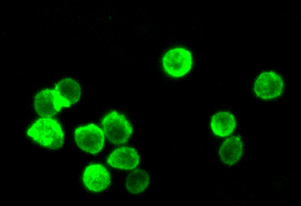
Cell Navigator 细胞膜染色试剂盒是美国AAT Bioquest生产的用于染色细胞膜的试剂盒,Cell Navigator 细胞质膜染色试剂盒可快速,均匀地标记质膜,而没有凝集素表现出的细胞类型差异。 它可以用作HCS(高含量筛选)的分割工具,以及对细胞质膜进行染色以用于标准荧光显微镜检查。 试剂盒中使用的绿色荧光染料可固定但不能通透,因此不适用于还涉及通过抗体探测内部靶标的实验。金畔生物是AAT Bioquest的中国代理商,为您提供最优质的Cell Navigator 细胞膜染色试剂盒。
点击查看光谱
适用仪器
| 荧光显微镜 | |
| 激发: | FITC滤波片 |
| 发射: | FITC滤波片 |
| 推荐孔板: | 黑色透明孔板或载玻片 |
产品说明书
样品实验方案
简要概述
1.在生长培养基中准备细胞
2.准备并向细胞添加Cellpaint Green工作溶液
3.在37℃下孵育5至20分钟
4.使用FITC滤光片组读取荧光强度
溶液配制
1.储备溶液配制
所有未使用的储备溶液应分为一次性使用的等分试样,并在制备后储存在-20°C下。 避免重复冻融循环。
Cellpaint 绿色原液(500X):
将100 uL DMSO(组分C)添加到Cellpaint Green(组分A)小瓶中,制成500X储备溶液。 注意:20 µL Cellpaint Green 500X储备溶液足以用于一个96孔板。 如果管密封严密,可以将未使用的Cellpaint Green 500X储备溶液分装并在≤-20ºC下保存1个月。 避光并避免重复的冻融循环。
2.工作溶液配制
Cellpaint 绿色工作溶液(1X):
将20 uL的500X储备溶液添加到10 mL的测定缓冲液(组分B)中,并充分混合。 注意:我们建议在使用前使工作溶液新鲜。
点击查看细胞制备方案
样品示例及操作
2.除去每个孔中的工作溶液。 用生理缓冲液(例如HHBS,DPBS或您选择的缓冲液)洗涤细胞3次,并替换为HHBS。
3.可选:染色后固定细胞。 用4%甲醛固定细胞15-30分钟。 用生理缓冲液洗涤细胞三遍。
4.使用带有FITC滤波片组的荧光显微镜观察细胞中的荧光信号。
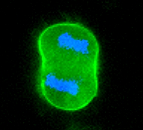
图1.在玻片上,Cellpaint Green与Hoechst 33342共染色的HL-60细胞的荧光图像。 使用荧光显微镜,使用带有FITC滤镜的60X油镜对细胞成像。 由于来自细胞边界上方和下方的膜的所有荧光信号均被不加选择地捕获,因此图像显得更加刺破和扩散。 与常规的落射荧光成像相比,共聚焦荧光成像比常规的落射荧光显微镜更灵敏,可以为您提供更多的控制。 如果可以使用共聚焦显微镜,则可能会获得清晰的细胞膜成像。
参考文献
Matrix Rigidity-Dependent Regulation of Ca(2+) at Plasma Membrane Microdomains by FAK Visualized by Fluorescence Resonance Energy Transfer
Authors: T. J. Kim
Journal: Adv Sci (Weinh) (2019): 1801290
Revealing the Raft Domain Organization in the Plasma Membrane by Single-Molecule Imaging of Fluorescent Ganglioside Analogs
Authors: K. G. N. Suzuki
Journal: Methods Enzymol (2018): 267-282
A single fluorescent probe enables clearly discriminating and simultaneously imaging liquid-ordered and liquid-disordered microdomains in plasma membrane of living cells
Authors: M. Tian
Journal: Biomaterials (2017): 46-56
A fluorescent cholesterol analogue for observation of free cholesterol in the plasma membrane of live cells
Authors: Y. Ogawa
Journal: Anal Biochem (2016): 49-55
Determination of Dynamics of Plant Plasma Membrane Proteins with Fluorescence Recovery and Raster Image Correlation Spectroscopy
Authors: M. Lankova
Journal: Microsc Microanal (2016): 290-9
Single-molecule Super-resolution Imaging of Phosphatidylinositol 4,5-bisphosphate in the Plasma Membrane with Novel Fluorescent Probes
Authors: C. Ji
Journal: J Vis Exp (2016): se name=”22682.enl” path=”C:UsersaatbiDropbox (AAT Bioquest)Website Working FilesProduct References22682.enl”>22682.enlEndNote4417Ji, C.Lou, X.Department of Neuroscience, University of Wisconsin-Madison. Department of Neuroscience, University of
Subcellular Fate of a Fluorescent Cholesterol-Poly(ethylene glycol) Conjugate: An Excellent Plasma Membrane Imaging Reagent
Authors: X. Chen
Journal: Langmuir (2016): 10126-10135
Fluorescent protein-based biosensors to visualize signal transduction beneath the plasma membrane
Authors: Y. Fujioka
Journal: Anal Sci (2015): 267-74
Imaging sub-plasma membrane cAMP dynamics with fluorescent translocation reporters
Authors: A. Tengholm
Journal: Methods Mol Biol (2015): 85-101
Role of the nucleocapsid domain in HIV-1 Gag oligomerization and trafficking to the plasma membrane: a fluorescence lifetime imaging microscopy investigation
Authors: S. E. El Meshri
Journal: J Mol Biol (2015): 1480-1494
相关产品
| 产品名称 | 货号 |
| Cell Navigator 细胞膜染色试剂盒 红色荧光 | Cat#22681 |
| Cell Navigator 细胞膜染色试剂盒 橙色荧光 | Cat#22680 |